1I06 | pdb_00001i06

CRYSTAL STRUCTURE OF MOUSE MAJOR URINARY PROTEIN (MUP-I) COMPLEXED WITH SEC-BUTYL-THIAZOLINE


Experimental Data Snapshot

  • Method: X-RAY DIFFRACTION
  • Resolution: 1.90 Å
  • R-Value Free: 
    0.247 (Depositor), 0.252 (DCC) 
  • R-Value Work: 
    0.217 (Depositor), 0.221 (DCC) 
  • R-Value Observed: 
    0.217 (Depositor) 

Starting Model: experimental
View more details

wwPDB Validation 3D Report Full Report

Validation slider image for 1I06

This is version 1.3 of the entry. See complete history

Literature

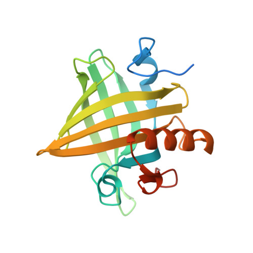
Structural Basis of Pheromone Binding to Mouse Major Urinary Protein (MUP-I)

Timm, D.E.Baker, L.J.Mueller, H.Zidek, L.Novotny, M.V.

(2001) Protein Sci 10: 997-1004

  • DOI: https://doi.org/10.1110/ps.52201
  • Primary Citation Related Structures: 
    1I04, 1I05, 1I06

  • PubMed Abstract: 

    The mouse major urinary proteins are pheromone-binding proteins that function as carriers of volatile effectors of mouse physiology and behavior. Crystal structures of recombinant mouse major urinary protein-I (MUP-I) complexed with the synthetic pheromones, 2-sec-butyl-4,5-dihydrothiazole and 6-hydroxy-6-methyl-3-heptanone, have been determined at high resolution. The purification of MUP-I from mouse liver and a high-resolution structure of the natural isolate are also reported. These results show the binding of 6-hydroxy-6-methyl-3-heptanone to MUP-I, unambiguously define ligand orientations for two pheromones within the MUP-I binding site, and suggest how different chemical classes of pheromones can be accommodated within the MUP-I beta-barrel.


  • Organizational Affiliation
    • Department of Biochemistry, Indiana University, Indianapolis, Indiana 46202, USA. dtimm@iupui.edu

Macromolecule Content 

  • Total Structure Weight: 21.27 kDa 
  • Atom Count: 1,310 
  • Modeled Residue Count: 156 
  • Deposited Residue Count: 180 
  • Unique protein chains: 1

Macromolecules

Find similar proteins by:|  3D Structure
Entity ID: 1
MoleculeChains  Sequence LengthOrganismDetailsImage
MAJOR URINARY PROTEIN I180Mus musculusMutation(s): 0 
UniProt
Find proteins for P02762 (Mus musculus)
Explore P02762 
Go to UniProtKB:  P02762
Entity Groups
Sequence Clusters30% Identity50% Identity70% Identity90% Identity95% Identity100% Identity
UniProt GroupP02762
Sequence Annotations
Expand
Reference Sequence

Experimental Data & Validation

Experimental Data

  • Method: X-RAY DIFFRACTION
  • Resolution: 1.90 Å
  • R-Value Free:  0.247 (Depositor), 0.252 (DCC) 
  • R-Value Work:  0.217 (Depositor), 0.221 (DCC) 
  • R-Value Observed: 0.217 (Depositor) 
Space Group: P 43 21 2
Unit Cell:
Length ( Å )Angle ( ˚ )
a = 57.033α = 90
b = 57.033β = 90
c = 108.976γ = 90
Software Package:
Software NamePurpose
DENZOdata reduction
SCALEPACKdata scaling
CNSrefinement
CNSphasing

Structure Validation

View Full Validation Report


Entry History 

Deposition Data

Revision History  (Full details and data files)

  • Version 1.0: 2001-02-14
    Type: Initial release
  • Version 1.1: 2008-04-27
    Changes: Version format compliance
  • Version 1.2: 2011-07-13
    Changes: Version format compliance
  • Version 1.3: 2023-08-09
    Changes: Data collection, Database references, Derived calculations, Refinement description