1HSB | pdb_00001hsb

DIFFERENT LENGTH PEPTIDES BIND TO HLA-AW68 SIMILARLY AT THEIR ENDS BUT BULGE OUT IN THE MIDDLE


Experimental Data Snapshot

  • Method: X-RAY DIFFRACTION
  • Resolution: 1.90 Å
  • R-Value Work: 
    0.220 (Depositor) 
  • R-Value Observed: 
    0.220 (Depositor) 

wwPDB Validation 3D Report Full Report

Validation slider image for 1HSB

This is version 1.4 of the entry. See complete history

Literature

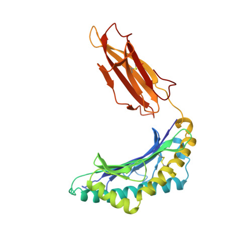
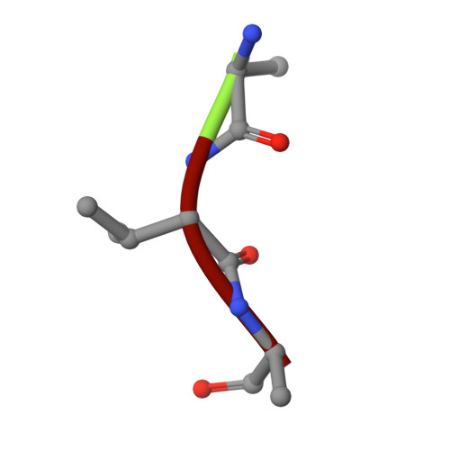
Different length peptides bind to HLA-Aw68 similarly at their ends but bulge out in the middle.

Guo, H.C.Jardetzky, T.S.Garrett, T.P.Lane, W.S.Strominger, J.L.Wiley, D.C.

(1992) Nature 360: 364-366

  • DOI: https://doi.org/10.1038/360364a0
  • Primary Citation Related Structures: 
    1HSB

  • PubMed Abstract: 

    We report here the determination and refinement to 1.9 A resolution by X-ray cryo-crystallography the structure of HLA-Aw68. The averaged image from the collection of bound, endogenous peptides clearly shows the atomic structure at the first three and last two amino acids in the peptides but no connected electron density in between. This suggests that bound peptides, held at both ends, take alternative pathways and could be of different lengths by bulging out in the middle. Peptides eluted from HLA-Aw68 include peptides of 9, 10 and 11 amino acids, a direct indication of the length heterogeneity of tightly bound peptides. Peptide sequencing shows relatively conserved 'anchor' residues at position 2 and the carboxy-terminal residue. Conserved binding sites for the peptide N and C termini at the ends of the class I major histocompatibility complex binding groove are apparently dominant in producing the long half-lives of peptide binding and the peptide-dependent stabilization of the class I molecule's structure.


  • Organizational Affiliation
    • Department of Biochemistry and Molecular Biology, Harvard University, Cambridge, Massachusetts 02138.

Macromolecule Content 

  • Total Structure Weight: 43.42 kDa 
  • Atom Count: 3,327 
  • Modeled Residue Count: 372 
  • Deposited Residue Count: 372 
  • Unique protein chains: 3

Macromolecules

Find similar proteins by:|  3D Structure
Entity ID: 1
MoleculeChains  Sequence LengthOrganismDetailsImage
MHC class I antigen270Homo sapiensMutation(s): 0 
UniProt & NIH Common Fund Data Resources
Find proteins for P04439 (Homo sapiens)
Explore P04439 
Go to UniProtKB:  P04439
PHAROS:  P04439
GTEx:  ENSG00000206503 
Entity Groups
Sequence Clusters30% Identity50% Identity70% Identity90% Identity95% Identity100% Identity
UniProt GroupP04439
Sequence Annotations
Expand
Reference Sequence
Find similar proteins by:|  3D Structure
Entity ID: 2
MoleculeChains  Sequence LengthOrganismDetailsImage
Beta-2-microglobulin99Homo sapiensMutation(s): 0 
UniProt & NIH Common Fund Data Resources
Find proteins for P61769 (Homo sapiens)
Explore P61769 
Go to UniProtKB:  P61769
PHAROS:  P61769
GTEx:  ENSG00000166710 
Entity Groups
Sequence Clusters30% Identity50% Identity70% Identity90% Identity95% Identity100% Identity
UniProt GroupP61769
Sequence Annotations
Expand
Reference Sequence
Find similar proteins by:  Sequence   |   3D Structure  
Entity ID: 3
MoleculeChains  Sequence LengthOrganismDetailsImage
BOUND PEPTIDE FRAGMENT3N/AMutation(s): 0 

Small Molecules

Ligands 2 Unique
IDChains Name / Formula / InChI Key2D Diagram3D Interactions
ARG

Query on ARG


Download:Ideal Coordinates CCD File
E [auth A]ARGININE
C6 H15 N4 O2
ODKSFYDXXFIFQN-BYPYZUCNSA-O
ALA

Query on ALA


Download:Ideal Coordinates CCD File
D [auth A]ALANINE
C3 H7 N O2
QNAYBMKLOCPYGJ-REOHCLBHSA-N

Experimental Data & Validation

Experimental Data

  • Method: X-RAY DIFFRACTION
  • Resolution: 1.90 Å
  • R-Value Work:  0.220 (Depositor) 
  • R-Value Observed: 0.220 (Depositor) 
Space Group: P 21 21 21
Unit Cell:
Length ( Å )Angle ( ˚ )
a = 59.43α = 90
b = 78.469β = 90
c = 111.904γ = 90
Software Package:
Software NamePurpose
X-PLORmodel building
X-PLORrefinement
X-PLORphasing

Structure Validation

View Full Validation Report


Entry History 

Deposition Data

Revision History  (Full details and data files)

  • Version 1.0: 1993-10-31
    Type: Initial release
  • Version 1.1: 2008-03-24
    Changes: Version format compliance
  • Version 1.2: 2011-07-13
    Changes: Version format compliance
  • Version 1.3: 2024-06-19
    Changes: Data collection, Database references, Derived calculations, Other, Source and taxonomy, Structure summary
  • Version 1.4: 2024-10-30
    Changes: Structure summary