1HRP

CRYSTAL STRUCTURE OF HUMAN CHORIONIC GONADOTROPIN


Experimental Data Snapshot

  • Method: X-RAY DIFFRACTION
  • Resolution: 3.00 Å
  • R-Value Free: 0.314 
  • R-Value Work: 0.218 
  • R-Value Observed: 0.218 

wwPDB Validation   3D Report Full Report


This is version 2.1 of the entry. See complete history


Literature

Crystal structure of human chorionic gonadotropin.

Lapthorn, A.J.Harris, D.C.Littlejohn, A.Lustbader, J.W.Canfield, R.E.Machin, K.J.Morgan, F.J.Isaacs, N.W.

(1994) Nature 369: 455-461

  • DOI: https://doi.org/10.1038/369455a0
  • Primary Citation of Related Structures:  
    1HRP

  • PubMed Abstract: 

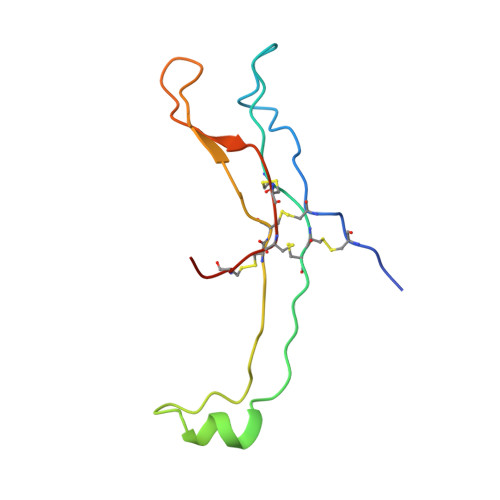
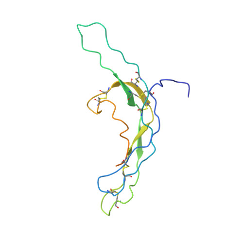
    The three-dimensional structure of human chorionic gonadotropin shows that each of its two different subunits has a similar topology, with three disulphide bonds forming a cystine knot. This same folding motif is found in some protein growth factors. The heterodimer is stabilized by a segment of the beta-subunit which wraps around the alpha-subunit and is covalently linked like a seat belt by the disulphide Cys 26-Cys 110. This extraordinary feature appears to be essential not only for the association of these heterodimers but also for receptor binding by the glycoprotein hormones.


  • Organizational Affiliation

    Department of Chemistry, University of Glasgow, UK.


Macromolecules
Find similar proteins by:  (by identity cutoff)  |  3D Structure
Entity ID: 1
MoleculeChains Sequence LengthOrganismDetailsImage
HUMAN CHORIONIC GONADOTROPIN92Homo sapiensMutation(s): 0 
UniProt & NIH Common Fund Data Resources
Find proteins for P01215 (Homo sapiens)
Explore P01215 
Go to UniProtKB:  P01215
PHAROS:  P01215
GTEx:  ENSG00000135346 
Entity Groups  
Sequence Clusters30% Identity50% Identity70% Identity90% Identity95% Identity100% Identity
UniProt GroupP01215
Glycosylation
Glycosylation Sites: 2Go to GlyGen: P01215-1
Sequence Annotations
Expand
  • Reference Sequence
Find similar proteins by:  (by identity cutoff)  |  3D Structure
Entity ID: 2
MoleculeChains Sequence LengthOrganismDetailsImage
HUMAN CHORIONIC GONADOTROPIN145Homo sapiensMutation(s): 0 
Gene Names: CGB3CGB5CGB8
UniProt & NIH Common Fund Data Resources
Find proteins for P0DN86 (Homo sapiens)
Explore P0DN86 
Go to UniProtKB:  P0DN86
PHAROS:  P0DN86
Entity Groups  
Sequence Clusters30% Identity50% Identity70% Identity90% Identity95% Identity100% Identity
UniProt GroupP0DN86
Glycosylation
Glycosylation Sites: 2Go to GlyGen: P0DN86-1
Sequence Annotations
Expand
  • Reference Sequence
Oligosaccharides

Help

Entity ID: 3
MoleculeChains Length2D Diagram Glycosylation3D Interactions
2-acetamido-2-deoxy-beta-D-glucopyranose-(1-4)-2-acetamido-2-deoxy-beta-D-glucopyranose
C, D
2N-Glycosylation
Glycosylation Resources
GlyTouCan:  G42666HT
GlyCosmos:  G42666HT
GlyGen:  G42666HT
Small Molecules
Ligands 1 Unique
IDChains Name / Formula / InChI Key2D Diagram3D Interactions
NAG
Query on NAG

Download Ideal Coordinates CCD File 
E [auth B],
F [auth B]
2-acetamido-2-deoxy-beta-D-glucopyranose
C8 H15 N O6
OVRNDRQMDRJTHS-FMDGEEDCSA-N
Experimental Data & Validation

Experimental Data

  • Method: X-RAY DIFFRACTION
  • Resolution: 3.00 Å
  • R-Value Free: 0.314 
  • R-Value Work: 0.218 
  • R-Value Observed: 0.218 
  • Space Group: P 65 2 2
Unit Cell:
Length ( Å )Angle ( ˚ )
a = 88.68α = 90
b = 88.68β = 90
c = 177.24γ = 120
Software Package:
Software NamePurpose
X-PLORmodel building
X-PLORrefinement
X-PLORphasing

Structure Validation

View Full Validation Report


Entry History 

Deposition Data

Revision History  (Full details and data files)

  • Version 1.0: 1994-11-01
    Type: Initial release
  • Version 1.1: 2008-03-03
    Changes: Version format compliance
  • Version 1.2: 2011-07-13
    Changes: Non-polymer description, Version format compliance
  • Version 2.0: 2020-07-29
    Type: Remediation
    Reason: Carbohydrate remediation
    Changes: Atomic model, Data collection, Database references, Derived calculations, Other, Structure summary
  • Version 2.1: 2024-10-30
    Changes: Data collection, Database references, Structure summary