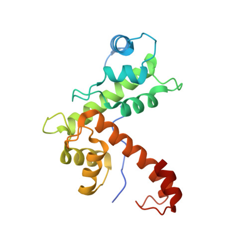
Structure of apoptosis-linked protein ALG-2: insights into Ca2+-induced changes in penta-EF-hand proteins.
Jia, J., Tarabykina, S., Hansen, C., Berchtold, M., Cygler, M.(2001) Structure 9: 267-275
- PubMed: 11525164 Search on PubMed
- DOI: https://doi.org/10.1016/s0969-2126(01)00585-8
- Primary Citation Related Structures:
1HQV - PubMed Abstract:
The Ca2+ binding apoptosis-linked gene-2 (ALG-2) protein acts as a proapoptotic factor in a variety of cell lines and is required either downstream or independently of caspases for apoptosis to occur. ALG-2 belongs to the penta-EF-hand (PEF) protein family and has two high-affinity and one low-affinity Ca2+ binding sites. Like other PEF proteins, its N terminus contains a Gly/Pro-rich segment. Ca2+ binding is required for the interaction with the target protein, ALG-2 interacting protein 1 (AIP1). We present the 2.3 A resolution crystal structure of Ca2+-Ioaded des1-20ALG-2 (aa 21-191), which was obtained by limited proteolysis of recombinant ALG-2 with elastase. The molecule contains eight alpha helices that fold into five EF-hands, and, similar to other members of this protein family, the molecule forms dimers. Ca2+ ions bind to EF1, EF3, and, surprisingly, to EF5. In the related proteins calpain and grancalcin, the EF5 does not bind Ca2+ and is thought to primarily facilitate dimerization. Most importantly, the conformation of des1-20ALG-2 is significantly different from that of calpain and grancalcin. This difference can be described as a rigid body rotation of EF1-2 relative to EF4-5 and the dimer interface, with a hinge within the EF3 loop. An electron density, which is interpreted as a hydrophobic Gly/Pro-rich decapeptide that is possibly derived from the cleaved N terminus, was found in a hydrophobic cleft between these two halves of the molecule. A different relative orientation of the N- and C-terminal halves of des1-20ALG-2 in the presence of Ca2+ and the peptide as compared to other Ca2+loaded PEF proteins changes substantially the shape of the molecule, exposing a hydrophobic patch on the surface for peptide binding and a large cleft near the dimer interface. We postulate that the binding of a Gly/ Pro-rich peptide in the presence of Ca2+ induces a conformational rearrangement in ALG-2, and that this mechanism is common to other PEF proteins.
- Biotechnology Research Institute, National Research Council of Canada, Montreal, Quebec.
Organizational Affiliation: