1HP5 | pdb_00001hp5

STREPTOMYCES PLICATUS BETA-N-ACETYLHEXOSAMINIDASE COMPLEXED WITH INTERMEDIATE ANALOUGE NAG-THIAZOLINE


Experimental Data Snapshot

  • Method: X-RAY DIFFRACTION
  • Resolution: 2.10 Å
  • R-Value Free: 
    0.222 (Depositor) 
  • R-Value Work: 
    0.197 (Depositor), 0.215 (DCC) 
  • R-Value Observed: 
    0.197 (Depositor) 

Starting Model: experimental
View more details

wwPDB Validation 3D Report Full Report

Validation slider image for 1HP5

Ligand Structure Quality Assessment 


This is version 2.1 of the entry. See complete history

Literature

Crystallographic evidence for substrate-assisted catalysis in a bacterial beta-hexosaminidase.

Mark, B.L.Vocadlo, D.J.Knapp, S.Triggs-Raine, B.L.Withers, S.G.James, M.N.

(2001) J Biological Chem 276: 10330-10337

  • DOI: https://doi.org/10.1074/jbc.M011067200
  • Primary Citation Related Structures: 
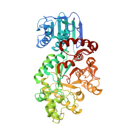
    1HP4, 1HP5

  • PubMed Abstract: 

    beta-Hexosaminidase, a family 20 glycosyl hydrolase, catalyzes the removal of beta-1,4-linked N-acetylhexosamine residues from oligosaccharides and their conjugates. Heritable deficiency of this enzyme results in various forms of GalNAc-beta(1,4)-[N-acetylneuraminic acid (2,3)]-Gal-beta(1,4)-Glc-ceramide gangliosidosis, including Tay-Sachs disease. We have determined the x-ray crystal structure of a beta-hexosaminidase from Streptomyces plicatus to 2.2 A resolution (Protein Data Bank code ). beta-Hexosaminidases are believed to use a substrate-assisted catalytic mechanism that generates a cyclic oxazolinium ion intermediate. We have solved and refined a complex between the cyclic intermediate analogue N-acetylglucosamine-thiazoline and beta-hexosaminidase from S. plicatus to 2.1 A resolution (Protein Data Bank code ). Difference Fourier analysis revealed the pyranose ring of N-acetylglucosamine-thiazoline bound in the enzyme active site with a conformation close to that of a (4)C(1) chair. A tryptophan-lined hydrophobic pocket envelopes the thiazoline ring, protecting it from solvolysis at the iminium ion carbon. Within this pocket, Tyr(393) and Asp(313) appear important for positioning the 2-acetamido group of the substrate for nucleophilic attack at the anomeric center and for dispersing the positive charge distributed into the oxazolinium ring upon cyclization. This complex provides decisive structural evidence for substrate-assisted catalysis and the formation of a covalent, cyclic intermediate in family 20 beta-hexosaminidases.


  • Organizational Affiliation
    • Medical Research Council Group in Protein Structure and Function, Department of Biochemistry, University of Alberta, Edmonton, Alberta T6G 2H7, Canada.

Macromolecule Content 

  • Total Structure Weight: 56.73 kDa 
  • Atom Count: 4,168 
  • Modeled Residue Count: 499 
  • Deposited Residue Count: 512 
  • Unique protein chains: 1

Macromolecules

Find similar proteins by:|  3D Structure
Entity ID: 1
MoleculeChains  Sequence LengthOrganismDetailsImage
BETA-N-ACETYLHEXOSAMINIDASE512Streptomyces plicatusMutation(s): 0 
EC: 3.2.1.52
UniProt
Find proteins for O85361 (Streptomyces plicatus)
Explore O85361 
Go to UniProtKB:  O85361
Entity Groups
Sequence Clusters30% Identity50% Identity70% Identity90% Identity95% Identity100% Identity
UniProt GroupO85361
Sequence Annotations
Expand
Reference Sequence

Small Molecules

Ligands 4 Unique
IDChains Name / Formula / InChI Key2D Diagram3D Interactions
NGT

Query on NGT


Download:Ideal Coordinates CCD File
F [auth A]3AR,5R,6S,7R,7AR-5-HYDROXYMETHYL-2-METHYL-5,6,7,7A-TETRAHYDRO-3AH-PYRANO[3,2-D]THIAZOLE-6,7-DIOL
C8 H13 N O4 S
DRHXTSWSUAJOJZ-FMDGEEDCSA-N
SO4

Query on SO4


Download:Ideal Coordinates CCD File
E [auth A]SULFATE ION
O4 S
QAOWNCQODCNURD-UHFFFAOYSA-L
GOL

Query on GOL


Download:Ideal Coordinates CCD File
G [auth A],
H [auth A]
GLYCEROL
C3 H8 O3
PEDCQBHIVMGVHV-UHFFFAOYSA-N
CL

Query on CL


Download:Ideal Coordinates CCD File
B [auth A],
C [auth A],
D [auth A]
CHLORIDE ION
Cl
VEXZGXHMUGYJMC-UHFFFAOYSA-M

Experimental Data & Validation

Experimental Data

  • Method: X-RAY DIFFRACTION
  • Resolution: 2.10 Å
  • R-Value Free:  0.222 (Depositor) 
  • R-Value Work:  0.197 (Depositor), 0.215 (DCC) 
  • R-Value Observed: 0.197 (Depositor) 
Space Group: P 61 2 2
Unit Cell:
Length ( Å )Angle ( ˚ )
a = 133.621α = 90
b = 133.621β = 90
c = 174.194γ = 120
Software Package:
Software NamePurpose
DENZOdata reduction
SCALEPACKdata scaling
CNSrefinement
CNSphasing

Structure Validation

View Full Validation Report


Ligand Structure Quality Assessment 


Entry History 

Deposition Data

  • Released Date: 2001-04-04 
  • Deposition Author(s): Mark, B.L.

Revision History  (Full details and data files)

  • Version 1.0: 2001-04-04
    Type: Initial release
  • Version 1.1: 2008-04-27
    Changes: Version format compliance
  • Version 1.2: 2011-07-13
    Changes: Derived calculations, Version format compliance
  • Version 2.0: 2023-08-09
    Changes: Atomic model, Data collection, Database references, Derived calculations, Refinement description
  • Version 2.1: 2024-10-16
    Changes: Structure summary