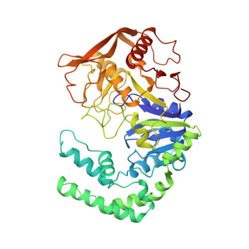
Refined crystal structures of guanine nucleotide complexes of adenylosuccinate synthetase from Escherichia coli.
Poland, B.W., Hou, Z., Bruns, C., Fromm, H.J., Honzatko, R.B.(1996) J Biol Chem 271: 15407-15413
- PubMed: 8663109
- DOI: https://doi.org/10.1074/jbc.271.26.15407
- Primary Citation of Related Structures:
1HON, 1HOO, 1HOP - PubMed Abstract:
Structures of adenylosuccinate synthetase from Escherichia coli complexed with guanosine-5'-(beta,gamma-imido) triphosphate and guanosine-5'-(beta,gamma-methylene)triphosphate in the presence and the absence of Mg2+ have been refined to R-factors below 0.2 against data to a nominal resolution of 2.7 A. Asp333 of the synthetase hydrogen bonds to the exocyclic 2-amino and endocyclic N1 groups of the guanine nucleotide base, whereas the hydroxyl of Ser414 and the backbone amide of Lys331 hydrogen bond to the 6-oxo position. The side chains of Lys331 and Pro417 pack against opposite faces of the guanine nucleotide base. The synthetase recognizes neither the N7 position of guanine nucleotides nor the ribose group. Electron density for the guanine-5'-(beta,gamma-imido) triphosphate complex is consistent with a mixture of the triphosphate nucleoside and its hydrolyzed diphosphate nucleoside bound to the active site. The base, ribose, and alpha-phosphate positions overlap, but the beta-phosphates occupy different binding sites. The binding of guanosine-5'-(beta,gamma-methylene)triphosphate to the active site is comparable with that of guanosine-5'-(beta, gamma-imido)triphosphate. No electron density, however, for the corresponding diphosphate nucleoside is observed. In addition, electron density for bound Mg2+ is absent in these nucleotide complexes. The guanine nucleotide complexes of the synthetase are compared with complexes of other GTP-binding proteins and to a preliminary structure of the complex of GDP, IMP, Mg2+, and succinate with the synthetase. The enzyme, under conditions reported here, does not undergo a conformational change in response to the binding of guanine nucleotides, and minimally IMP and/or Mg2+ must be present in order to facilitate the complete recognition of the guanine nucleotide by the synthetase.
Organizational Affiliation:
Department of Biochemistry and Biophysics, Iowa State University, Ames, Iowa 50011, USA.


