1HJ9 | pdb_00001hj9

Atomic resolution structures of trypsin provide insight into structural radiation damage


Experimental Data Snapshot

  • Method: X-RAY DIFFRACTION
  • Resolution: 0.95 Å
  • R-Value Free: 
    0.140 (Depositor), 0.115 (DCC) 
  • R-Value Work: 
    0.122 (DCC) 
  • R-Value Observed: 
    0.117 (Depositor) 

wwPDB Validation 3D Report Full Report

Validation slider image for 1HJ9

This is version 2.1 of the entry. See complete history

Literature

Atomic Resolution Structure of Trypsin Provide Insight Into Structural Radiation Damage

Leiros, H.-K.S.Mcsweeney, S.M.Smalas, A.O.

(2001) Acta Crystallogr D Biol Crystallogr 57: 488

  • DOI: https://doi.org/10.1107/s0907444901000646
  • Primary Citation Related Structures: 
    1HJ8, 1HJ9

  • PubMed Abstract: 

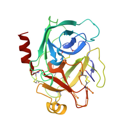
    Radiation damage is an inherent problem in protein X-ray crystallography and the process has recently been shown to be highly specific, exhibiting features such as cleavage of disulfide bonds, decarboxylation of acidic residues, increase in atomic B factors and increase in unit-cell volume. Reported here are two trypsin structures at atomic resolution (1.00 and 0.95 A), the data for which were collected at a third-generation synchrotron (ESRF) at two different beamlines. Both trypsin structures exhibit broken disulfide bonds; in particular, the bond from Cys191 to Cys220 is very sensitive to synchrotron radiation. The data set collected at the most intense beamline (ID14-EH4) shows increased structural radiation damage in terms of lower occupancies for cysteine residues, more breakage in the six disulfide bonds and more alternate conformations. It appears that high intensity and not only the total X-ray dose is most harmful to protein crystals.


  • Organizational Affiliation
    • Protein Crystallography Group, Department of Chemistry, Faculty of Science, University of Tromsø, N-9037 Tromsø, Norway.

Macromolecule Content 

  • Total Structure Weight: 23.84 kDa 
  • Atom Count: 1,965 
  • Modeled Residue Count: 223 
  • Deposited Residue Count: 223 
  • Unique protein chains: 1

Macromolecules

Find similar proteins by:|  3D Structure
Entity ID: 1
MoleculeChains  Sequence LengthOrganismDetailsImage
BETA-TRYPSIN223Bos taurusMutation(s): 0 
EC: 3.4.21.4
UniProt
Find proteins for P00760 (Bos taurus)
Explore P00760 
Go to UniProtKB:  P00760
Entity Groups
Sequence Clusters30% Identity50% Identity70% Identity90% Identity95% Identity100% Identity
UniProt GroupP00760
Sequence Annotations
Expand
Reference Sequence

Small Molecules

Ligands 4 Unique
IDChains Name / Formula / InChI Key2D Diagram3D Interactions
SO4

Query on SO4


Download:Ideal Coordinates CCD File
C [auth A],
D [auth A],
E [auth A]
SULFATE ION
O4 S
QAOWNCQODCNURD-UHFFFAOYSA-L
ANL

Query on ANL


Download:Ideal Coordinates CCD File
B [auth A]ANILINE
C6 H7 N
PAYRUJLWNCNPSJ-UHFFFAOYSA-N
GOL

Query on GOL


Download:Ideal Coordinates CCD File
F [auth A]GLYCEROL
C3 H8 O3
PEDCQBHIVMGVHV-UHFFFAOYSA-N
CA

Query on CA


Download:Ideal Coordinates CCD File
G [auth A]CALCIUM ION
Ca
BHPQYMZQTOCNFJ-UHFFFAOYSA-N

Experimental Data & Validation

Experimental Data

  • Method: X-RAY DIFFRACTION
  • Resolution: 0.95 Å
  • R-Value Free:  0.140 (Depositor), 0.115 (DCC) 
  • R-Value Work:  0.122 (DCC) 
  • R-Value Observed: 0.117 (Depositor) 
Space Group: P 21 21 21
Unit Cell:
Length ( Å )Angle ( ˚ )
a = 54.055α = 90
b = 56.812β = 90
c = 66.282γ = 90
Software Package:
Software NamePurpose
SHELXL-97refinement
DENZOdata reduction
SCALAdata scaling

Structure Validation

View Full Validation Report


Entry History 

Deposition Data

Revision History  (Full details and data files)

  • Version 1.0: 2002-01-04
    Type: Initial release
  • Version 1.1: 2011-05-08
    Changes: Version format compliance
  • Version 1.2: 2011-07-13
    Changes: Version format compliance
  • Version 2.0: 2025-04-09
    Changes: Advisory, Atomic model, Data collection, Database references, Derived calculations, Other, Structure summary
  • Version 2.1: 2025-10-01
    Changes: Derived calculations, Structure summary