1HIW | pdb_00001hiw

TRIMERIC HIV-1 MATRIX PROTEIN


Experimental Data Snapshot

  • Method: X-RAY DIFFRACTION
  • Resolution: 2.30 Å
  • R-Value Free: 
    0.332 (Depositor) 
  • R-Value Work: 
    0.259 (Depositor), 0.248 (DCC) 
  • R-Value Observed: 
    0.259 (Depositor) 

wwPDB Validation 3D Report Full Report

Validation slider image for 1HIW

This is version 1.3 of the entry. See complete history

Literature

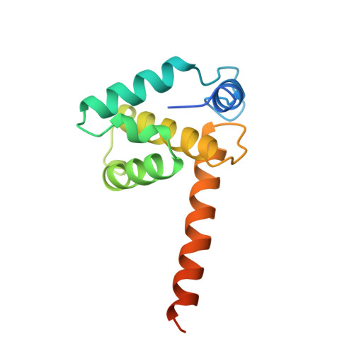
Crystal structures of the trimeric human immunodeficiency virus type 1 matrix protein: implications for membrane association and assembly.

Hill, C.P.Worthylake, D.Bancroft, D.P.Christensen, A.M.Sundquist, W.I.

(1996) Proc Natl Acad Sci U S A 93: 3099-3104

  • DOI: https://doi.org/10.1073/pnas.93.7.3099
  • Primary Citation Related Structures: 
    1HIW

  • PubMed Abstract: 

    The human immunodeficiency virus type 1 (HIV-1) matrix protein forms a structural shell associated with the inner viral membrane and performs other essential functions throughout the viral life cycle. The crystal structure of the HIV-1 matrix protein, determined at 2.3 angstrom resolution, reveals that individual matrix molecules are composed of five major helices capped by a three-stranded mixed beta-sheet. Unexpectedly, the protein assembles into a trimer in three different crystal lattices, burying 1880 angstrom2 of accessible surface area at the trimer interfaces. Trimerization appears to create a large, bipartite membrane binding surface in which exposed basic residues could cooperate with the N-terminal myristoyl groups to anchor the protein on the acidic inner membrane of the virus.


  • Organizational Affiliation
    • Department of Biochemistry, University of Utah, Salt Lake City, 84132, USA.

Macromolecule Content 

  • Total Structure Weight: 90.79 kDa 
  • Atom Count: 5,348 
  • Modeled Residue Count: 658 
  • Deposited Residue Count: 798 
  • Unique protein chains: 1

Macromolecules

Find similar proteins by:|  3D Structure
Entity ID: 1
MoleculeChains  Sequence LengthOrganismDetailsImage
HIV-1 MATRIX PROTEIN133Human immunodeficiency virus 1Mutation(s): 0 
UniProt
Find proteins for P12497 (Human immunodeficiency virus type 1 group M subtype B (isolate NY5))
Explore P12497 
Go to UniProtKB:  P12497
Entity Groups
Sequence Clusters30% Identity50% Identity70% Identity90% Identity95% Identity100% Identity
UniProt GroupP12497
Sequence Annotations
Expand
Reference Sequence

Experimental Data & Validation

Experimental Data

  • Method: X-RAY DIFFRACTION
  • Resolution: 2.30 Å
  • R-Value Free:  0.332 (Depositor) 
  • R-Value Work:  0.259 (Depositor), 0.248 (DCC) 
  • R-Value Observed: 0.259 (Depositor) 
Space Group: P 1 21 1
Unit Cell:
Length ( Å )Angle ( ˚ )
a = 64.423α = 90
b = 91.163β = 102.91
c = 73.997γ = 90
Software Package:
Software NamePurpose
X-PLORmodel building
X-PLORrefinement
DENZOdata reduction
SCALEPACKdata scaling
X-PLORphasing

Structure Validation

View Full Validation Report


Entry History 

Deposition Data

Revision History  (Full details and data files)

  • Version 1.0: 1996-10-14
    Type: Initial release
  • Version 1.1: 2008-03-24
    Changes: Version format compliance
  • Version 1.2: 2011-07-13
    Changes: Version format compliance
  • Version 1.3: 2024-02-07
    Changes: Data collection, Database references, Derived calculations, Other