Comparative analysis of the X-ray structures of HIV-1 and HIV-2 proteases in complex with CGP 53820, a novel pseudosymmetric inhibitor.
Priestle, J.P., Fassler, A., Rosel, J., Tintelnot-Blomley, M., Strop, P., Grutter, M.G.(1995) Structure 3: 381-389
- PubMed: 7613867
- DOI: https://doi.org/10.1016/s0969-2126(01)00169-1
- Primary Citation of Related Structures:
1HIH, 1HII - PubMed Abstract:
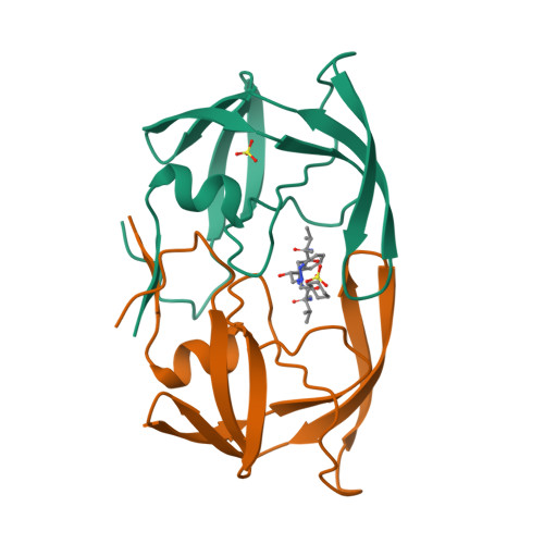
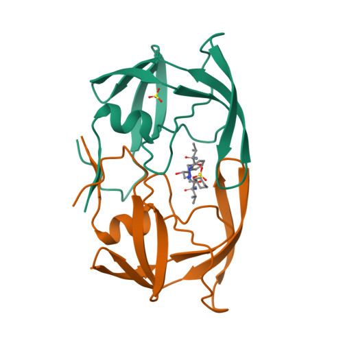
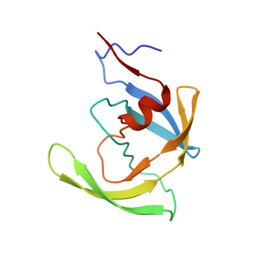
The human immunodeficiency virus (HIV) is the causative agent of acquired immunodeficiency syndrome (AIDS). Two subtypes of the virus, HIV-1 and HIV-2, have been characterized. The protease enzymes from these two subtypes, which are aspartic acid proteases and have been found to be essential for maturation of the infectious particle, share about 50% sequence identity. Differences in substrate and inhibitor binding between these enzymes have been previously reported. We report the X-ray crystal structures of both HIV-1 and HIV-2 proteases each in complex with the pseudosymmetric inhibitor, CGP 53820, to 2.2 A and 2.3 A, respectively. In both structures, the entire enzyme and inhibitor could be located. The structures confirmed earlier modeling studies. Differences between the CGP 53820 inhibitory binding constants for the two enzymes could be correlated with structural differences. Minor sequence changes in subsites at the active site can explain some of the observed differences in substrate and inhibitor binding between the two enzymes. The information gained from this investigation may help in the design of equipotent HIV-1/HIV-2 protease inhibitors.
Organizational Affiliation:
Department of Core Drug Discovery Technologies, Pharma Research, Ciba-Geigy Ltd., Basel, Switzerland.


