1HG4 | pdb_00001hg4

Ultraspiracle ligand binding domain from Drosophila melanogaster


Experimental Data Snapshot

  • Method: X-RAY DIFFRACTION
  • Resolution: 2.40 Å
  • R-Value Free: 
    0.284 (Depositor) 
  • R-Value Work: 
    0.248 (Depositor) 
  • R-Value Observed: 
    0.248 (Depositor) 

Starting Model: experimental
View more details

wwPDB Validation 3D Report Full Report

Validation slider image for 1HG4

This is version 1.2 of the entry. See complete history

Literature

The Structure of the Ultraspiracle Ligand-Binding Domain Reveals a Nuclear Receptor Locked in an Inactive Conformation

Clayton, G.M.Peak-Chew, S.Y.Evans, R.M.Schwabe, J.W.R.

(2001) Proc Natl Acad Sci U S A 98: 1549

  • DOI: https://doi.org/10.1073/pnas.98.4.1549
  • Primary Citation Related Structures: 
    1HG4

  • PubMed Abstract: 

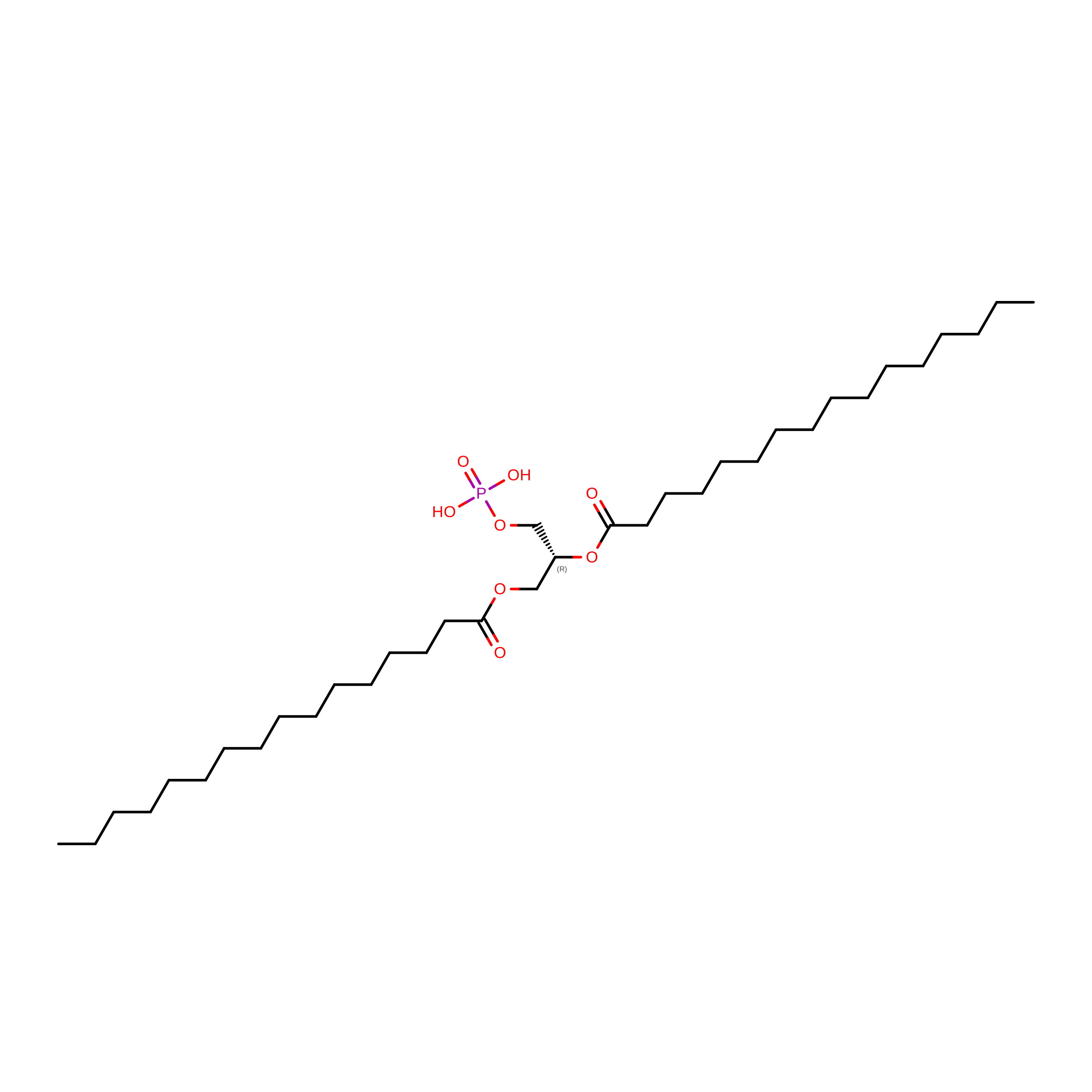
    Ultraspiracle (USP) is the invertebrate homologue of the mammalian retinoid X receptor (RXR). RXR plays a uniquely important role in differentiation, development, and homeostasis through its ability to serve as a heterodimeric partner to many other nuclear receptors. RXR is able to influence the activity of its partner receptors through the action of the ligand 9-cis retinoic acid. In contrast to RXR, USP has no known high-affinity ligand and is thought to be a silent component in the heterodimeric complex with partner receptors such as the ecdysone receptor. Here we report the 2.4-A crystal structure of the USP ligand-binding domain. The structure shows that a conserved sequence motif found in dipteran and lepidopteran USPs, but not in mammalian RXRs, serves to lock USP in an inactive conformation. It also shows that USP has a large hydrophobic cavity, implying that there is almost certainly a natural ligand for USP. This cavity is larger than that seen previously for most other nuclear receptors. Intriguingly, this cavity has partial occupancy by a bound lipid, which is likely to resemble the natural ligand for USP.


  • Organizational Affiliation
    • Medical Research Council, Laboratory of Molecular Biology, Cambridge CB2 2QH, United Kingdom.

Macromolecule Content 

  • Total Structure Weight: 192.02 kDa 
  • Atom Count: 11,854 
  • Modeled Residue Count: 1,436 
  • Deposited Residue Count: 1,674 
  • Unique protein chains: 1

Macromolecules

Find similar proteins by:|  3D Structure
Entity ID: 1
MoleculeChains  Sequence LengthOrganismDetailsImage
ULTRASPIRACLE
A, B, C, D, E
A, B, C, D, E, F
279Drosophila melanogasterMutation(s): 0 
UniProt
Find proteins for P20153 (Drosophila melanogaster)
Explore P20153 
Go to UniProtKB:  P20153
Entity Groups
Sequence Clusters30% Identity50% Identity70% Identity90% Identity95% Identity100% Identity
UniProt GroupP20153
Sequence Annotations
Expand
Reference Sequence

Experimental Data & Validation

Experimental Data

  • Method: X-RAY DIFFRACTION
  • Resolution: 2.40 Å
  • R-Value Free:  0.284 (Depositor) 
  • R-Value Work:  0.248 (Depositor) 
  • R-Value Observed: 0.248 (Depositor) 
Space Group: P 1
Unit Cell:
Length ( Å )Angle ( ˚ )
a = 37.98α = 85.57
b = 86.19β = 85.94
c = 137γ = 83.09
Software Package:
Software NamePurpose
CNSrefinement
MOSFLMdata reduction
CNSphasing

Structure Validation

View Full Validation Report


Entry History 

Deposition Data

Revision History  (Full details and data files)

  • Version 1.0: 2001-02-23
    Type: Initial release
  • Version 1.1: 2015-02-11
    Changes: Database references, Derived calculations, Non-polymer description, Other, Refinement description, Source and taxonomy, Structure summary, Version format compliance
  • Version 1.2: 2023-12-13
    Changes: Data collection, Database references, Derived calculations, Refinement description