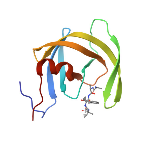
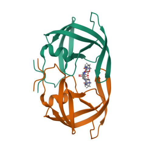
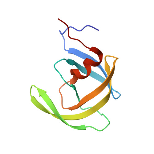
1HEG

The crystal structures at 2.2 angstroms resolution of hydroxyethylene-based inhibitors bound to human immunodeficiency virus type 1 protease show that the inhibitors are present in two distinct orientations


Experimental Data Snapshot

  • Method: X-RAY DIFFRACTION
  • Resolution: 2.20 Å
  • R-Value Work: 0.186 
  • R-Value Observed: 0.186 

wwPDB Validation   3D Report Full Report


This is version 1.6 of the entry. See complete history


Literature

The crystal structures at 2.2-A resolution of hydroxyethylene-based inhibitors bound to human immunodeficiency virus type 1 protease show that the inhibitors are present in two distinct orientations.

Murthy, K.H.Winborne, E.L.Minnich, M.D.Culp, J.S.Debouck, C.

(1992) J Biol Chem 267: 22770-22778

  • Primary Citation of Related Structures:  
    1HEF, 1HEG

  • PubMed Abstract: 

    As part of a structure-based drug design program directed against enzyme targets in the human immunodeficiency virus (HIV), we have determined the three-dimensional structures of the HIV type 1 protease complexed with two hydroxyethylene-based inhibitors. The inhibitors (SKF 107457 and SKF 108738) are hexapeptide substrate analogues with the scissile bond being replaced by a hydroxyethylene isostere. The structures were determined using x-ray diffraction data to 2.2 A measured at the Cornell High Energy Synchrotron Source on hexagonal crystals of each of the complexes. The structures have been extensively refined using a reciprocal space least-squares method to conventional crystallographic R factors of 0.186 and 0.159, respectively. The protein structure differs from that in the unliganded state of the enzyme and is most similar to that of the structure of the other reported (Jaskolski, M., Tomasselli, A. G., Sawyer, T. K., Staples, D. G., Heinrikson, R. L., Schneider, J., Kent, S. B. H., and Wlodawer, A. (1990) Biochemistry 29, 5889-5907) hydroxyethylene-based inhibitor complex. Unlike in that structure, however, the inhibitors are observed, in the present crystal structures, in two equally abundant orientations that are a consequence of the homodimeric nature of the enzyme coupled with the asymmetric structures of the inhibitors. Although the differences between the two inhibitors used in the present study are confined to the P1' site, the van der Waals interactions made by the inhibitor atoms with the amino acid residues in the protein differ throughout the structures of the inhibitors.


  • Organizational Affiliation

    Department of Macro Molecular Sciences, SmithKline Beecham Pharmaceuticals, King of Prussia, Pennsylvania 19406.


Macromolecules
Find similar proteins by:  (by identity cutoff)  |  3D Structure
Entity ID: 1
MoleculeChains Sequence LengthOrganismDetailsImage
HIV-1 PROTEASEA [auth E]99Human immunodeficiency virus 1Mutation(s): 0 
UniProt
Find proteins for P03366 (Human immunodeficiency virus type 1 group M subtype B (isolate BH10))
Explore P03366 
Go to UniProtKB:  P03366
Entity Groups  
Sequence Clusters30% Identity50% Identity70% Identity90% Identity95% Identity100% Identity
UniProt GroupP03366
Sequence Annotations
Expand
  • Reference Sequence
Small Molecules
Ligands 1 Unique
IDChains Name / Formula / InChI Key2D Diagram3D Interactions
PSI
Query on PSI

Download Ideal Coordinates CCD File 
B [auth E]methyl N-{(4S,5S)-5-[(L-alanyl-L-alanyl)amino]-4-hydroxy-6-phenylhexanoyl}-L-valyl-L-valinate
C29 H47 N5 O7
IUDCAKKZLXFOQA-QJAPXLAMSA-N
Biologically Interesting Molecules (External Reference) 1 Unique
Experimental Data & Validation

Experimental Data

  • Method: X-RAY DIFFRACTION
  • Resolution: 2.20 Å
  • R-Value Work: 0.186 
  • R-Value Observed: 0.186 
  • Space Group: P 61 2 2
Unit Cell:
Length ( Å )Angle ( ˚ )
a = 64.14α = 90
b = 64.14β = 90
c = 84.24γ = 120
Software Package:
Software NamePurpose
PROLSQrefinement

Structure Validation

View Full Validation Report


Entry History 

Deposition Data

Revision History  (Full details and data files)

  • Version 1.0: 1994-05-31
    Type: Initial release
  • Version 1.1: 2008-05-22
    Changes: Version format compliance
  • Version 1.2: 2011-07-13
    Changes: Atomic model, Database references, Derived calculations, Non-polymer description, Structure summary, Version format compliance
  • Version 1.3: 2012-12-12
    Changes: Other
  • Version 1.4: 2017-11-29
    Changes: Derived calculations, Other
  • Version 1.5: 2024-02-07
    Changes: Data collection, Database references, Derived calculations
  • Version 1.6: 2024-03-13
    Changes: Source and taxonomy, Structure summary