1HE8 | pdb_00001he8

Ras G12V - PI 3-kinase gamma complex


Experimental Data Snapshot

  • Method: X-RAY DIFFRACTION
  • Resolution: 3.00 Å
  • R-Value Free: 
    0.280 (Depositor), 0.274 (DCC) 
  • R-Value Work: 
    0.212 (Depositor), 0.210 (DCC) 
  • R-Value Observed: 
    0.212 (Depositor) 

Starting Models: experimental
View more details

wwPDB Validation 3D Report Full Report

Validation slider image for 1HE8

Ligand Structure Quality Assessment 


This is version 1.5 of the entry. See complete history

Literature

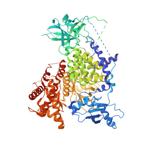
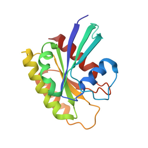
Crystal Structure and Functional Analysis of Ras Binding to its Effector Phosphoinositide 3-Kinase Gamma

Pacold, M.E.Suire, S.Perisic, O.Lara-Gonzalez, S.Davis, C.T.Walker, E.H.Hawkins, P.T.Stephens, L.Eccleston, J.F.Williams, R.L.

(2000) Cell 103: 931

  • DOI: https://doi.org/10.1016/s0092-8674(00)00196-3
  • Primary Citation Related Structures: 
    1HE8

  • PubMed Abstract: 

    Ras activation of phosphoinositide 3-kinase (PI3K) is important for survival of transformed cells. We find that PI3Kgamma is strongly and directly activated by H-Ras G12V in vivo or by GTPgammaS-loaded H-Ras in vitro. We have determined a crystal structure of a PI3Kgamma/Ras.GMPPNP complex. A critical loop in the Ras binding domain positions Ras so that it uses its switch I and switch II regions to bind PI3Kgamma. Mutagenesis shows that interactions with both regions are essential for binding PI3Kgamma. Ras also forms a direct contact with the PI3Kgamma catalytic domain. These unique Ras/PI3Kgamma interactions are likely to be shared by PI3Kalpha. The complex with Ras shows a change in the PI3K conformation that may represent an allosteric component of Ras activation.


  • Organizational Affiliation
    • MRC Laboratory of Molecular Biology Hills Road CB2 2QH, Cambridge, United Kingdom.

Macromolecule Content 

  • Total Structure Weight: 130.09 kDa 
  • Atom Count: 7,434 
  • Modeled Residue Count: 915 
  • Deposited Residue Count: 1,131 
  • Unique protein chains: 2

Macromolecules

Find similar proteins by:|  3D Structure
Entity ID: 1
MoleculeChains  Sequence LengthOrganismDetailsImage
PHOSPHATIDYLINOSITOL 3-KINASE CATALYTIC SUBUNIT, GAMMA ISOFORM965Homo sapiensMutation(s): 2 
EC: 2.7.1.137 (PDB Primary Data), 2.7.1.154 (UniProt), 2.7.11.1 (UniProt), 2.7.1.153 (UniProt)
UniProt & NIH Common Fund Data Resources
Find proteins for P48736 (Homo sapiens)
Explore P48736 
Go to UniProtKB:  P48736
PHAROS:  P48736
GTEx:  ENSG00000105851 
Entity Groups
Sequence Clusters30% Identity50% Identity70% Identity90% Identity95% Identity100% Identity
UniProt GroupP48736
Sequence Annotations
Expand
Reference Sequence
Find similar proteins by:|  3D Structure
Entity ID: 2
MoleculeChains  Sequence LengthOrganismDetailsImage
TRANSFORMING PROTEIN P21/H-RAS-1166Homo sapiensMutation(s): 1 
EC: 3.6.5.2
UniProt & NIH Common Fund Data Resources
Find proteins for P01112 (Homo sapiens)
Explore P01112 
Go to UniProtKB:  P01112
PHAROS:  P01112
GTEx:  ENSG00000174775 
Entity Groups
Sequence Clusters30% Identity50% Identity70% Identity90% Identity95% Identity100% Identity
UniProt GroupP01112
Sequence Annotations
Expand
Reference Sequence

Small Molecules

Ligands 2 Unique
IDChains Name / Formula / InChI Key2D Diagram3D Interactions
GNP

Query on GNP


Download:Ideal Coordinates CCD File
D [auth B]PHOSPHOAMINOPHOSPHONIC ACID-GUANYLATE ESTER
C10 H17 N6 O13 P3
UQABYHGXWYXDTK-UUOKFMHZSA-N
MG

Query on MG


Download:Ideal Coordinates CCD File
C [auth B]MAGNESIUM ION
Mg
JLVVSXFLKOJNIY-UHFFFAOYSA-N

Experimental Data & Validation

Experimental Data

  • Method: X-RAY DIFFRACTION
  • Resolution: 3.00 Å
  • R-Value Free:  0.280 (Depositor), 0.274 (DCC) 
  • R-Value Work:  0.212 (Depositor), 0.210 (DCC) 
  • R-Value Observed: 0.212 (Depositor) 
Space Group: P 31 2 1
Unit Cell:
Length ( Å )Angle ( ˚ )
a = 113.635α = 90
b = 113.635β = 90
c = 183.881γ = 120
Software Package:
Software NamePurpose
CNSrefinement
MOSFLMdata reduction
SCALAdata scaling
CNSphasing

Structure Validation

View Full Validation Report


Ligand Structure Quality Assessment 


Entry History 

Deposition Data

Revision History  (Full details and data files)

  • Version 1.0: 2001-01-08
    Type: Initial release
  • Version 1.1: 2011-05-08
    Changes: Version format compliance
  • Version 1.2: 2011-07-13
    Changes: Version format compliance
  • Version 1.3: 2017-07-12
    Changes: Derived calculations
  • Version 1.4: 2019-04-03
    Changes: Data collection, Source and taxonomy
  • Version 1.5: 2023-12-13
    Changes: Data collection, Database references, Derived calculations, Other, Refinement description