1HDT | pdb_00001hdt

STRUCTURE OF A RETRO-BINDING PEPTIDE INHIBITOR COMPLEXED WITH HUMAN ALPHA-THROMBIN


Experimental Data Snapshot

  • Method: X-RAY DIFFRACTION
  • Resolution: 2.60 Å
  • R-Value Work: 
    0.204 (Depositor) 
  • R-Value Observed: 
    0.204 (Depositor) 

wwPDB Validation 3D Report Full Report

Validation slider image for 1HDT

This is version 1.5 of the entry. See complete history

Literature

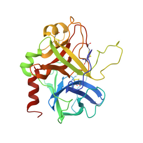
Structure of a retro-binding peptide inhibitor complexed with human alpha-thrombin.

Tabernero, L.Chang, C.Y.Ohringer, S.L.Lau, W.F.Iwanowicz, E.J.Han, W.C.Wang, T.C.Seiler, S.M.Roberts, D.G.Sack, J.S.

(1995) J Mol Biology 246: 14-20

  • DOI: https://doi.org/10.1006/jmbi.1994.0060
  • Primary Citation Related Structures: 
    1HDT

  • PubMed Abstract: 

    The crystallographic structure of the ternary complex between human alpha-thrombin, hirugen and the peptidyl inhibitor Phe-alloThr-Phe-O-CH3, which is acylated at its N terminus with 4-guanidino butanoic acid (BMS-183507), has been determined at 2.6 A resolution. The structure reveals a unique "retro-binding" mode for this tripeptide active site inhibitor. The inhibitor binds with its alkyl-guanidine moiety in the primary specificity pocket and its two phenyl rings occupying the hydrophobic proximal and distal pockets of the thrombin active site. In this arrangement the backbone of the tripeptide forms a parallel beta-strand to the thrombin main-chain at the binding site. This is opposite to the orientation of the natural substrate, fibrinogen, and all the small active site-directed thrombin inhibitors whose bound structures have been previously reported. BMS-183507 is the first synthetic inhibitor proved to bind in a retro-binding fashion to thrombin, in a fashion similar to that of the N-terminal residues of the natural inhibitor hirudin. Furthermore, this new potent thrombin inhibitor (Ki = 17.2 nM) is selective for thrombin over other serine proteases tested and may be a template to be considered in designing hirudin-based thrombin inhibitors with interactions at the specificity pocket.


  • Organizational Affiliation
    • Department of Macromolecular Crystallography, Bristol-Myers Squibb Pharmaceutical Research Institute, Princeton, NJ 08543-4000.

Macromolecule Content 

  • Total Structure Weight: 35.67 kDa 
  • Atom Count: 2,597 
  • Modeled Residue Count: 303 
  • Deposited Residue Count: 304 
  • Unique protein chains: 3

Macromolecules

Find similar proteins by:|  3D Structure
Entity ID: 1
MoleculeChains  Sequence LengthOrganismDetailsImage
ALPHA-THROMBINA [auth L]33Homo sapiensMutation(s): 0 
EC: 3.4.21.5
UniProt & NIH Common Fund Data Resources
Find proteins for P00734 (Homo sapiens)
Explore P00734 
Go to UniProtKB:  P00734
PHAROS:  P00734
GTEx:  ENSG00000180210 
Entity Groups
Sequence Clusters30% Identity50% Identity70% Identity90% Identity95% Identity100% Identity
UniProt GroupP00734
Sequence Annotations
Expand
Reference Sequence
Find similar proteins by:|  3D Structure
Entity ID: 2
MoleculeChains  Sequence LengthOrganismDetailsImage
ALPHA-THROMBINB [auth H]259Homo sapiensMutation(s): 0 
EC: 3.4.21.5
UniProt & NIH Common Fund Data Resources
Find proteins for P00734 (Homo sapiens)
Explore P00734 
Go to UniProtKB:  P00734
PHAROS:  P00734
GTEx:  ENSG00000180210 
Entity Groups
Sequence Clusters30% Identity50% Identity70% Identity90% Identity95% Identity100% Identity
UniProt GroupP00734
Sequence Annotations
Expand
Reference Sequence
Find similar proteins by:  Sequence   |   3D Structure  
Entity ID: 3
MoleculeChains  Sequence LengthOrganismDetailsImage
HIRUGEN PEPTIDEC [auth P]12Hirudo medicinalisMutation(s): 0 
UniProt
Find proteins for P28509 (Hirudo medicinalis)
Explore P28509 
Go to UniProtKB:  P28509
Entity Groups
Sequence Clusters30% Identity50% Identity70% Identity90% Identity95% Identity100% Identity
UniProt GroupP28509
Sequence Annotations
Expand
Reference Sequence

Small Molecules

Ligands 1 Unique
IDChains Name / Formula / InChI Key2D Diagram3D Interactions
0E7

Query on 0E7


Download:Ideal Coordinates CCD File
D [auth H]methyl N-(4-carbamimidamidobutanoyl)-L-phenylalanyl-L-allothreonyl-L-phenylalaninate
C28 H38 N6 O6
QKSIUCFYHKTUCL-CKLTXHEASA-N
Modified Residues  1 Unique
IDChains TypeFormula2D DiagramParent
TYS
Query on TYS
C [auth P]L-PEPTIDE LINKINGC9 H11 N O6 STYR

Biologically Interesting Molecules (External Reference) 

1 Unique

Experimental Data & Validation

Experimental Data

  • Method: X-RAY DIFFRACTION
  • Resolution: 2.60 Å
  • R-Value Work:  0.204 (Depositor) 
  • R-Value Observed: 0.204 (Depositor) 
Space Group: P 21 21 2
Unit Cell:
Length ( Å )Angle ( ˚ )
a = 108.97α = 90
b = 80.92β = 90
c = 46.17γ = 90
Software Package:
Software NamePurpose
X-PLORmodel building
X-PLORrefinement
X-PLORphasing

Structure Validation

View Full Validation Report


Entry History 

Deposition Data

Revision History  (Full details and data files)

  • Version 1.0: 1995-10-15
    Type: Initial release
  • Version 1.1: 2008-03-03
    Changes: Version format compliance
  • Version 1.2: 2011-07-13
    Changes: Atomic model, Database references, Derived calculations, Non-polymer description, Structure summary, Version format compliance
  • Version 1.3: 2012-12-12
    Changes: Other
  • Version 1.4: 2024-06-05
    Changes: Data collection, Database references, Derived calculations, Other
  • Version 1.5: 2024-10-16
    Changes: Structure summary