1HDG | pdb_00001hdg

THE CRYSTAL STRUCTURE OF HOLO-GLYCERALDEHYDE-3-PHOSPHATE DEHYDROGENASE FROM THE HYPERTHERMOPHILIC BACTERIUM THERMOTOGA MARITIMA AT 2.5 ANGSTROMS RESOLUTION


Experimental Data Snapshot

  • Method: X-RAY DIFFRACTION
  • Resolution: 2.50 Å
  • R-Value Work: 
    0.166 (Depositor) 
  • R-Value Observed: 
    0.166 (Depositor) 

wwPDB Validation 3D Report Full Report

Validation slider image for 1HDG

This is version 1.3 of the entry. See complete history

Literature

The crystal structure of holo-glyceraldehyde-3-phosphate dehydrogenase from the hyperthermophilic bacterium Thermotoga maritima at 2.5 A resolution.

Korndorfer, I.Steipe, B.Huber, R.Tomschy, A.Jaenicke, R.

(1995) J Mol Biology 246: 511-521

  • DOI: https://doi.org/10.1006/jmbi.1994.0103
  • Primary Citation Related Structures: 
    1HDG

  • PubMed Abstract: 

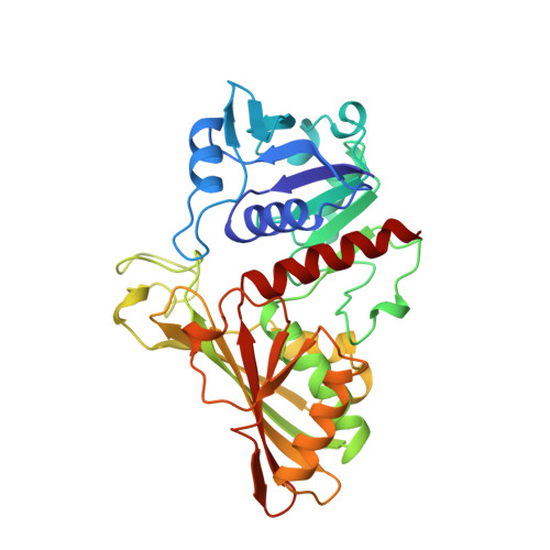
    The crystal structure of holo-glyceraldehyde-3-phosphate dehydrogenase from the hyperthermophile Thermotoga maritima was determined by Patterson search methods using the known structure of the Bacillus stearothermophilus enzyme. The structure was refined at a resolution of 2.5 A to an R-factor of 16.63% for 26289 reflections between 8.0 A an 2.5 A with F > 2 sigma(F). The crystallographic asymmetric unit contains two monomers related by approximate 2-fold symmetry and a tetramer is built up by crystallographic symmetry. The root-mean-square deviation of Ca positions of glyceraldehyde-3-phosphate dehydrogenase from T. maritima and B. stearothermophilus is 0.83 A in the NAD+ binding domains and smaller close to the cofactor. In contrast, the largest deviations in the catalytic domains are found at residues involved in coordination of sulphate ion SO4 339, which most likely marks the site of the attacking inorganic phosphate ion in catalysis. A large number of extra salt-bridges may be an important factor contributing to the high thermostability of this protein.


  • Organizational Affiliation
    • Max-Planck Institut für Biochemie, Martinsried, FRG.

Macromolecule Content 

  • Total Structure Weight: 74.39 kDa 
  • Atom Count: 5,422 
  • Modeled Residue Count: 664 
  • Deposited Residue Count: 664 
  • Unique protein chains: 1

Macromolecules

Find similar proteins by:|  3D Structure
Entity ID: 1
MoleculeChains  Sequence LengthOrganismDetailsImage
HOLO-D-GLYCERALDEHYDE-3-PHOSPHATE DEHYDROGENASEA [auth O],
B [auth Q]
332Thermotoga maritimaMutation(s): 0 
Gene Names: TN10
EC: 1.2.1.12
UniProt
Find proteins for P17721 (Thermotoga maritima (strain ATCC 43589 / DSM 3109 / JCM 10099 / NBRC 100826 / MSB8))
Explore P17721 
Go to UniProtKB:  P17721
Entity Groups
Sequence Clusters30% Identity50% Identity70% Identity90% Identity95% Identity100% Identity
UniProt GroupP17721
Sequence Annotations
Expand
Reference Sequence

Experimental Data & Validation

Experimental Data

  • Method: X-RAY DIFFRACTION
  • Resolution: 2.50 Å
  • R-Value Work:  0.166 (Depositor) 
  • R-Value Observed: 0.166 (Depositor) 
Space Group: P 61 2 2
Unit Cell:
Length ( Å )Angle ( ˚ )
a = 95.7α = 90
b = 95.7β = 90
c = 296.28γ = 120
Software Package:
Software NamePurpose
X-PLORmodel building
X-PLORrefinement
X-PLORphasing

Structure Validation

View Full Validation Report


Entry History 

Deposition Data

Revision History  (Full details and data files)

  • Version 1.0: 1995-03-31
    Type: Initial release
  • Version 1.1: 2008-03-21
    Changes: Version format compliance
  • Version 1.2: 2011-07-13
    Changes: Derived calculations, Version format compliance
  • Version 1.3: 2024-02-07
    Changes: Data collection, Database references, Derived calculations, Other