1H80 | pdb_00001h80

1,3-ALPHA-1,4-BETA-D-GALACTOSE-4-SULFATE- 3,6-ANHYDRO-D-GALACTOSE-2-SULFATE 4 GALACTOHYDROLASE


Experimental Data Snapshot

  • Method: X-RAY DIFFRACTION
  • Resolution: 1.60 Å
  • R-Value Free: 
    0.223 (Depositor), 0.226 (DCC) 
  • R-Value Work: 
    0.207 (Depositor), 0.210 (DCC) 
  • R-Value Observed: 
    0.207 (Depositor) 

wwPDB Validation 3D Report Full Report

Validation slider image for 1H80

This is version 1.4 of the entry. See complete history

Literature

The Iota-Carrageenase of Alteromonas Fortis. A Beta-Helix Fold-Containing Enzyme for the Degradation of a Highly Polyanionic Polysaccharide

Michel, G.Chantalat, L.Fanchon, E.Henrissat, B.Kloareg, B.Dideberg, O.

(2001) J Biological Chem 276: 40202

  • DOI: https://doi.org/10.1074/jbc.M100670200
  • Primary Citation Related Structures: 
    1H80

  • PubMed Abstract: 

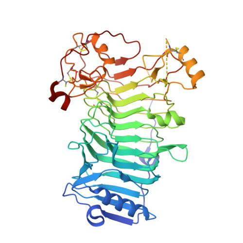
    Carrageenans are gel-forming hydrocolloids extracted from the cell walls of marine red algae. They consist of d-galactose residues bound by alternate alpha(1-->3) and beta(1-->4) linkages and substituted by one (kappa-carrageenan), two (iota-carrageenan), or three (lambda-carrageenan) sulfate-ester groups per disaccharide repeating unit. Both the kappa- and iota-carrageenan chains adopt ordered conformations leading to the formation of highly ordered aggregates of double-stranded helices. Several kappa-carrageenases and iota-carrageenases have been cloned from marine bacteria. Kappa-carrageenases belong to family 16 of the glycoside hydrolases, which essentially encompasses polysaccharidases specialized in the hydrolysis of the neutral polysaccharides such as agarose, laminarin, lichenan, and xyloglucan. In contrast, iota-carrageenases constitute a novel glycoside hydrolase structural family. We report here the crystal structure of Alteromonas fortis iota-carrageenase at 1.6 A resolution. The enzyme folds into a right-handed parallel beta-helix of 10 complete turns with two additional C-terminal domains. Glu(245), Asp(247), or Glu(310), in the cleft of the enzyme, are proposed as candidate catalytic residues. The protein contains one sodium and one chloride binding site and three calcium binding sites shown to be involved in stabilizing the enzyme structure.


  • Organizational Affiliation
    • Laboratoire de Cristallographie Macromoléculaire, Institut de Biologie Structurale Jean-Pierre Ebel, CNRS/Commissariat à l'Energie Atomique, 41, rue Jules Horowitz, 38027 Grenoble Cedex 1, France.

Macromolecule Content 

  • Total Structure Weight: 104.87 kDa 
  • Atom Count: 7,992 
  • Modeled Residue Count: 863 
  • Deposited Residue Count: 928 
  • Unique protein chains: 1

Macromolecules

Find similar proteins by:|  3D Structure
Entity ID: 1
MoleculeChains  Sequence LengthOrganismDetailsImage
IOTA-CARRAGEENASE
A, B
464Alteromonas macleodiiMutation(s): 0 
Gene Names: CGIA
EC: 3.2.1.157
UniProt
Find proteins for Q9F5I8 (Alteromonas macleodii)
Explore Q9F5I8 
Go to UniProtKB:  Q9F5I8
Entity Groups
Sequence Clusters30% Identity50% Identity70% Identity90% Identity95% Identity100% Identity
UniProt GroupQ9F5I8
Sequence Annotations
Expand
Reference Sequence

Small Molecules

Ligands 4 Unique
IDChains Name / Formula / InChI Key2D Diagram3D Interactions
GOL

Query on GOL


Download:Ideal Coordinates CCD File
J [auth A]
K [auth A]
L [auth A]
T [auth B]
U [auth B]
J [auth A],
K [auth A],
L [auth A],
T [auth B],
U [auth B],
V [auth B]
GLYCEROL
C3 H8 O3
PEDCQBHIVMGVHV-UHFFFAOYSA-N
CA

Query on CA


Download:Ideal Coordinates CCD File
C [auth A]
D [auth A]
E [auth A]
M [auth B]
N [auth B]
C [auth A],
D [auth A],
E [auth A],
M [auth B],
N [auth B],
O [auth B]
CALCIUM ION
Ca
BHPQYMZQTOCNFJ-UHFFFAOYSA-N
CL

Query on CL


Download:Ideal Coordinates CCD File
F [auth A],
P [auth B]
CHLORIDE ION
Cl
VEXZGXHMUGYJMC-UHFFFAOYSA-M
NA

Query on NA


Download:Ideal Coordinates CCD File
G [auth A]
H [auth A]
I [auth A]
Q [auth B]
R [auth B]
G [auth A],
H [auth A],
I [auth A],
Q [auth B],
R [auth B],
S [auth B]
SODIUM ION
Na
FKNQFGJONOIPTF-UHFFFAOYSA-N

Experimental Data & Validation

Experimental Data

  • Method: X-RAY DIFFRACTION
  • Resolution: 1.60 Å
  • R-Value Free:  0.223 (Depositor), 0.226 (DCC) 
  • R-Value Work:  0.207 (Depositor), 0.210 (DCC) 
  • R-Value Observed: 0.207 (Depositor) 
Space Group: P 1 21 1
Unit Cell:
Length ( Å )Angle ( ˚ )
a = 55.87α = 90
b = 90.07β = 93.53
c = 124.12γ = 90
Software Package:
Software NamePurpose
CNSrefinement
DENZOdata reduction
SCALEPACKdata scaling
SHARPphasing

Structure Validation

View Full Validation Report


Entry History 

Deposition Data

Revision History  (Full details and data files)

  • Version 1.0: 2001-11-27
    Type: Initial release
  • Version 1.1: 2011-05-08
    Changes: Version format compliance
  • Version 1.2: 2011-07-13
    Changes: Version format compliance
  • Version 1.3: 2018-10-24
    Changes: Advisory, Data collection, Source and taxonomy
  • Version 1.4: 2024-11-20
    Changes: Advisory, Data collection, Database references, Derived calculations, Other, Structure summary