1H6R | pdb_00001h6r

The oxidized state of a redox sensitive variant of green fluorescent protein


Experimental Data Snapshot

  • Method: X-RAY DIFFRACTION
  • Resolution: 1.50 Å
  • R-Value Free: 
    0.212 (Depositor) 
  • R-Value Work: 
    0.183 (Depositor), 0.179 (DCC) 
  • R-Value Observed: 
    0.183 (Depositor) 

Starting Model: experimental
View more details

wwPDB Validation 3D Report Full Report

Validation slider image for 1H6R

This is version 1.5 of the entry. See complete history

Literature

Shedding Light on Disulfide Bond Formation: Engineering a Redox Switch in Green Fluorescent Protein

Ostergaard, H.Henriksen, A.Hansen, F.G.Winther, J.R.

(2001) EMBO J 20: 5853

  • DOI: https://doi.org/10.1093/emboj/20.21.5853
  • Primary Citation Related Structures: 
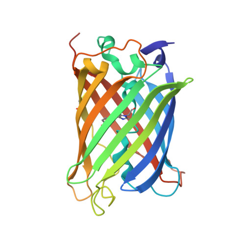
    1H6R

  • PubMed Abstract: 

    To visualize the formation of disulfide bonds in living cells, a pair of redox-active cysteines was introduced into the yellow fluorescent variant of green fluorescent protein. Formation of a disulfide bond between the two cysteines was fully reversible and resulted in a >2-fold decrease in the intrinsic fluorescence. Inter conversion between the two redox states could thus be followed in vitro as well as in vivo by non-invasive fluorimetric measurements. The 1.5 A crystal structure of the oxidized protein revealed a disulfide bond-induced distortion of the beta-barrel, as well as a structural reorganization of residues in the immediate chromophore environment. By combining this information with spectroscopic data, we propose a detailed mechanism accounting for the observed redox state-dependent fluorescence. The redox potential of the cysteine couple was found to be within the physiological range for redox-active cysteines. In the cytoplasm of Escherichia coli, the protein was a sensitive probe for the redox changes that occur upon disruption of the thioredoxin reductive pathway.


  • Organizational Affiliation
    • Section of Molecular Microbiology, BioCentrum-DTU, Technical University of Denmark, Building 301, DK-2800 Lyngby, Denmark.

Macromolecule Content 

  • Total Structure Weight: 80.9 kDa 
  • Atom Count: 6,447 
  • Modeled Residue Count: 687 
  • Deposited Residue Count: 708 
  • Unique protein chains: 1

Macromolecules

Find similar proteins by:|  3D Structure
Entity ID: 1
MoleculeChains  Sequence LengthOrganismDetailsImage
GREEN FLUORESCENT PROTEIN
A, B, C
236Aequorea victoriaMutation(s): 9 
UniProt
Find proteins for P42212 (Aequorea victoria)
Explore P42212 
Go to UniProtKB:  P42212
Entity Groups
Sequence Clusters30% Identity50% Identity70% Identity90% Identity95% Identity100% Identity
UniProt GroupP42212
Sequence Annotations
Expand
Reference Sequence

Small Molecules

Modified Residues  1 Unique
IDChains TypeFormula2D DiagramParent
PIA
Query on PIA
A, B, C
L-PEPTIDE LINKINGC14 H15 N3 O4ALA, TYR, GLY

Experimental Data & Validation

Experimental Data

  • Method: X-RAY DIFFRACTION
  • Resolution: 1.50 Å
  • R-Value Free:  0.212 (Depositor) 
  • R-Value Work:  0.183 (Depositor), 0.179 (DCC) 
  • R-Value Observed: 0.183 (Depositor) 
Space Group: P 21 21 21
Unit Cell:
Length ( Å )Angle ( ˚ )
a = 57.864α = 90
b = 93.915β = 90
c = 140.551γ = 90
Software Package:
Software NamePurpose
CNSrefinement
DENZOdata reduction
SCALEPACKdata scaling
AMoREphasing

Structure Validation

View Full Validation Report


Entry History 

Deposition Data

Revision History  (Full details and data files)

  • Version 1.0: 2001-11-15
    Type: Initial release
  • Version 1.1: 2011-05-08
    Changes: Version format compliance
  • Version 1.2: 2011-07-13
    Changes: Version format compliance
  • Version 1.3: 2018-01-17
    Changes: Data collection
  • Version 1.4: 2019-10-23
    Changes: Data collection, Database references, Derived calculations, Other
  • Version 1.5: 2023-12-13
    Changes: Data collection, Database references, Derived calculations, Refinement description