1H5Z | pdb_00001h5z

CYTOCHROME P450 14 ALPHA-STEROL DEMETHYLASE (CYP51) FROM MYCOBACTERIUM TUBERCULOSIS IN FERRIC LOW-SPIN STATE


Experimental Data Snapshot

  • Method: X-RAY DIFFRACTION
  • Resolution: 2.05 Å
  • R-Value Free: 
    0.236 (Depositor), 0.231 (DCC) 
  • R-Value Work: 
    0.185 (Depositor), 0.178 (DCC) 
  • R-Value Observed: 
    0.185 (Depositor) 

Starting Model: experimental
View more details

wwPDB Validation 3D Report Full Report

Validation slider image for 1H5Z

Ligand Structure Quality Assessment 


This is version 1.4 of the entry. See complete history

Literature

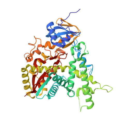
Estriol Bound and Ligand-Free Structures of Sterol 14Alpha-Demethylase.

Podust, L.M.Yermalitskaya, L.V.Lepesheva, G.I.Podust, V.N.Dalmasso, E.A.Waterman, M.R.

(2004) Structure 12: 1937

  • DOI: https://doi.org/10.1016/j.str.2004.08.009
  • Primary Citation Related Structures: 
    1H5Z, 1X8V

  • PubMed Abstract: 

    Sterol 14alpha-demethylases (CYP51) are essential enzymes in sterol biosynthesis in eukaryotes and drug targets in antifungal therapy. Here, we report CYP51 structures in ligand-free and estriol bound forms. Using estriol as a probe, we determined orientation of the substrate in the active site, elucidated protein contacts with the invariant 3beta-hydroxy group of a sterol, and identified F78 as a key discriminator between 4alpha-methylated and 4alpha,beta-dimethylated substrates. Analysis of CYP51 dynamics revealed that the C helix undergoes helix-coil transition upon binding and dissociation of a ligand. Loss of helical structure of the C helix in the ligand-free form results in an unprecedented opening of the substrate binding site. Upon binding of estriol, the BC loop loses contacts with molecular surface and tends to adopt a closed conformation. A mechanism for azole resistance in the yeast pathogen Candida albicans associated with mutations in the ERG11 gene encoding CYP51 is suggested based on CYP51 protein dynamics.


  • Organizational Affiliation
    • Department of Biochemistry, Vanderbilt University School of Medicine, Nashville, TN 37232, USA. larissa.m.podust@vanderbilt.edu

Macromolecule Content 

  • Total Structure Weight: 52.17 kDa 
  • Atom Count: 3,761 
  • Modeled Residue Count: 435 
  • Deposited Residue Count: 455 
  • Unique protein chains: 1

Macromolecules

Find similar proteins by:|  3D Structure
Entity ID: 1
MoleculeChains  Sequence LengthOrganismDetailsImage
CYTOCHROME P450 51455Mycobacterium tuberculosisMutation(s): 0 
Gene Names: CYP51RV0764CMTCY369.09C
EC: 1.14.13.70 (PDB Primary Data), 1.14.15.36 (UniProt)
UniProt
Find proteins for P9WPP9 (Mycobacterium tuberculosis (strain ATCC 25618 / H37Rv))
Explore P9WPP9 
Go to UniProtKB:  P9WPP9
Entity Groups
Sequence Clusters30% Identity50% Identity70% Identity90% Identity95% Identity100% Identity
UniProt GroupP9WPP9
Sequence Annotations
Expand
Reference Sequence

Small Molecules

Experimental Data & Validation

Experimental Data

  • Method: X-RAY DIFFRACTION
  • Resolution: 2.05 Å
  • R-Value Free:  0.236 (Depositor), 0.231 (DCC) 
  • R-Value Work:  0.185 (Depositor), 0.178 (DCC) 
  • R-Value Observed: 0.185 (Depositor) 
Space Group: P 21 21 21
Unit Cell:
Length ( Å )Angle ( ˚ )
a = 46.489α = 90
b = 84.827β = 90
c = 110.025γ = 90
Software Package:
Software NamePurpose
CNSrefinement
DENZOdata reduction
SCALEPACKdata scaling
CNSphasing

Structure Validation

View Full Validation Report


Ligand Structure Quality Assessment 


Entry History 

Deposition Data

Revision History  (Full details and data files)

  • Version 1.0: 2003-10-03
    Type: Initial release
  • Version 1.1: 2015-04-15
    Changes: Database references, Derived calculations, Non-polymer description, Other, Version format compliance
  • Version 1.2: 2015-08-05
    Changes: Derived calculations, Other, Source and taxonomy
  • Version 1.3: 2019-05-08
    Changes: Data collection, Experimental preparation, Other
  • Version 1.4: 2023-12-13
    Changes: Data collection, Database references, Derived calculations, Other, Refinement description