1H4R

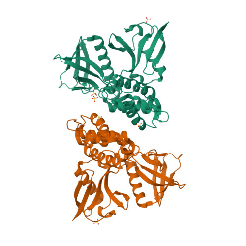
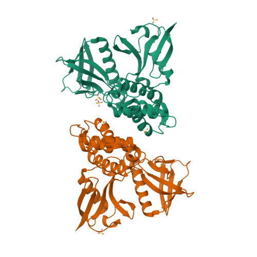
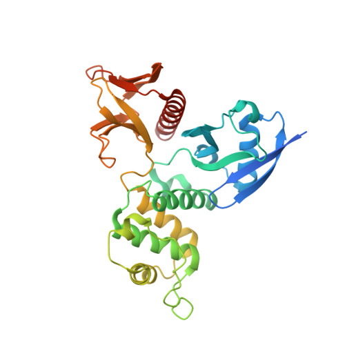
Crystal Structure of the FERM domain of Merlin, the Neurofibromatosis 2 Tumor Suppressor Protein.


Experimental Data Snapshot

  • Method: X-RAY DIFFRACTION
  • Resolution: 1.80 Å
  • R-Value Free: 0.227 
  • R-Value Work: 0.193 

Starting Model: experimental
View more details

wwPDB Validation   3D Report Full Report


This is version 1.4 of the entry. See complete history


Literature

The Structure of the Ferm Domain of Merlin, the Neurofibromatosis Type 2 Gene Product.

Kang, B.S.Cooper, D.R.Devedjiev, Y.Derewenda, U.Derewenda, Z.S.

(2002) Acta Crystallogr D Biol Crystallogr 58: 381

  • DOI: https://doi.org/10.1107/s0907444901021175
  • Primary Citation of Related Structures:  
    1H4R

  • PubMed Abstract: 

    Neurofibromatosis type 2 is an autosomal dominant disorder characterized by central nervous system tumors. The cause of the disease has been traced to mutations in the gene coding for a protein that is alternately called merlin or schwannomin and is a member of the ERM family (ezrin, radixin and moesin). The ERM proteins link the cytoskeleton to the cell membrane either directly through integral membrane proteins or indirectly through membrane-associated proteins. In this paper, the expression, purification, crystallization and crystal structure of the N-terminal domain of merlin are described. The crystals exhibit the symmetry of space group P2(1)2(1)2(1), with two molecules in the asymmetric unit. The recorded diffraction pattern extends to 1.8A resolution. The structure was solved by the molecular-replacement method and the model was refined to a conventional R value of 19.3% (R(free) = 22.7%). The N-terminal domain of merlin closely resembles those described for the corresponding domains in moesin and radixin and exhibits a cloverleaf architecture with three distinct subdomains. The structure allows a better rationalization of the impact of selected disease-causing mutations on the integrity of the protein.


  • Organizational Affiliation

    Department of Molecular Physiology and Biological Physics, University of Virginia, Charlottesville, Virginia 22908-0736, USA.


Macromolecules
Find similar proteins by:  (by identity cutoff)  |  3D Structure
Entity ID: 1
MoleculeChains Sequence LengthOrganismDetailsImage
MERLIN
A, B
314Homo sapiensMutation(s): 0 
UniProt & NIH Common Fund Data Resources
Find proteins for P35240 (Homo sapiens)
Explore P35240 
Go to UniProtKB:  P35240
PHAROS:  P35240
GTEx:  ENSG00000186575 
Entity Groups  
Sequence Clusters30% Identity50% Identity70% Identity90% Identity95% Identity100% Identity
UniProt GroupP35240
Sequence Annotations
Expand
  • Reference Sequence
Experimental Data & Validation

Experimental Data

  • Method: X-RAY DIFFRACTION
  • Resolution: 1.80 Å
  • R-Value Free: 0.227 
  • R-Value Work: 0.193 
  • Space Group: P 21 21 21
Unit Cell:
Length ( Å )Angle ( ˚ )
a = 87.018α = 90
b = 89.328β = 90
c = 96.764γ = 90
Software Package:
Software NamePurpose
REFMACrefinement
HKL-2000data reduction
HKL-2000data scaling
AMoREphasing

Structure Validation

View Full Validation Report


Entry History 

Deposition Data

Revision History  (Full details and data files)

  • Version 1.0: 2002-01-16
    Type: Initial release
  • Version 1.1: 2011-05-08
    Changes: Version format compliance
  • Version 1.2: 2011-07-13
    Changes: Version format compliance
  • Version 1.3: 2019-05-15
    Changes: Advisory, Data collection, Experimental preparation, Other
  • Version 1.4: 2023-12-13
    Changes: Advisory, Data collection, Database references, Other, Refinement description