1H1T | pdb_00001h1t

PHOSPHOPANTETHEINE ADENYLYLTRANSFERASE IN COMPLEX WITH Coenzyme A FROM ESCHERICHIA COLI


Experimental Data Snapshot

  • Method: X-RAY DIFFRACTION
  • Resolution: 1.78 Å
  • R-Value Free: 
    0.241 (Depositor) 
  • R-Value Work: 
    0.218 (Depositor) 
  • R-Value Observed: 
    0.218 (Depositor) 

Starting Model: experimental
View more details

wwPDB Validation 3D Report Full Report

Validation slider image for 1H1T

This is version 1.4 of the entry. See complete history

Literature

A Novel Adenylate Binding Site Confers Phosphopantetheine Adenylyltransferase Interactions with Coenzyme A

Izard, T.

(2003) J Bacteriol 185: 4074

  • DOI: https://doi.org/10.1128/JB.185.14.4074-4080.2003
  • Primary Citation Related Structures: 
    1H1T

  • PubMed Abstract: 

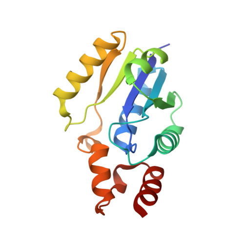
    Phosphopantetheine adenylyltransferase (PPAT) regulates the key penultimate step in the essential coenzyme A (CoA) biosynthetic pathway. PPAT catalyzes the reversible transfer of an adenylyl group from Mg(2+):ATP to 4'-phosphopantetheine to form 3'-dephospho-CoA (dPCoA) and pyrophosphate. The high-resolution crystal structure of PPAT complexed with CoA has been determined. Remarkably, CoA and the product dPCoA bind to the active site in distinct ways. Although the phosphate moiety within the phosphopantetheine arm overlaps, the pantetheine arm binds to the same pocket in two distinct conformations, and the adenylyl moieties of these two ligands have distinct binding sites. Moreover, the PPAT:CoA crystal structure confirms the asymmetry of binding to the two trimers within the hexameric enzyme. Specifically, the pantetheine arm of CoA bound to one protomer within the asymmetric unit displays the dPCoA-like conformation with the adenylyl moiety disordered, whereas CoA binds the twofold-related protomer in an ordered and unique fashion.


  • Organizational Affiliation
    • Department of Hematology-Oncology, St. Jude Children's Research Hospital, Memphis, Tennessee 38105-2794, USA. Tina.Izard@stjude.org

Macromolecule Content 

  • Total Structure Weight: 37.23 kDa 
  • Atom Count: 2,857 
  • Modeled Residue Count: 315 
  • Deposited Residue Count: 318 
  • Unique protein chains: 1

Macromolecules

Find similar proteins by:|  3D Structure
Entity ID: 1
MoleculeChains  Sequence LengthOrganismDetailsImage
PHOSPHOPANTETHEINE ADENYLYLTRANSFERASE
A, B
159Escherichia coliMutation(s): 0 
EC: 2.7.7.3
UniProt
Find proteins for P0A6I6 (Escherichia coli (strain K12))
Explore P0A6I6 
Go to UniProtKB:  P0A6I6
Entity Groups
Sequence Clusters30% Identity50% Identity70% Identity90% Identity95% Identity100% Identity
UniProt GroupP0A6I6
Sequence Annotations
Expand
Reference Sequence

Experimental Data & Validation

Experimental Data

  • Method: X-RAY DIFFRACTION
  • Resolution: 1.78 Å
  • R-Value Free:  0.241 (Depositor) 
  • R-Value Work:  0.218 (Depositor) 
  • R-Value Observed: 0.218 (Depositor) 
Space Group: I 2 3
Unit Cell:
Length ( Å )Angle ( ˚ )
a = 135.426α = 90
b = 135.426β = 90
c = 135.426γ = 90
Software Package:
Software NamePurpose
CNSrefinement
DENZOdata reduction
SCALEPACKdata scaling
CNSphasing

Structure Validation

View Full Validation Report


Entry History 

Deposition Data

  • Released Date: 2003-07-07 
  • Deposition Author(s): Izard, T.

Revision History  (Full details and data files)

  • Version 1.0: 2003-07-07
    Type: Initial release
  • Version 1.1: 2011-05-08
    Changes: Version format compliance
  • Version 1.2: 2011-07-13
    Changes: Version format compliance
  • Version 1.3: 2017-06-28
    Changes: Data collection
  • Version 1.4: 2023-12-13
    Changes: Data collection, Database references, Refinement description