1H1N | pdb_00001h1n

Atomic resolution structure of the major endoglucanase from Thermoascus aurantiacus


Experimental Data Snapshot

  • Method: X-RAY DIFFRACTION
  • Resolution: 1.12 Å
  • R-Value Free: 
    0.171 (Depositor), 0.164 (DCC) 
  • R-Value Work: 
    0.133 (DCC) 
  • R-Value Observed: 
    0.144 (Depositor) 

wwPDB Validation 3D Report Full Report

Validation slider image for 1H1N

This is version 1.6 of the entry. See complete history

Literature

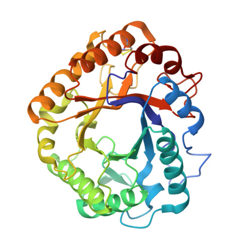
Atomic Resolution Structure of the Major Endoglucanase from Thermoascus Aurantiacus

Van Petegem, F.Vandenberghe, I.Bhat, M.K.Van Beeumen, J.

(2002) Biochem Biophys Res Commun 296: 161

  • DOI: https://doi.org/10.1016/s0006-291x(02)00775-1
  • Primary Citation Related Structures: 
    1H1N

  • PubMed Abstract: 

    The crystal structure of the major endoglucanase from the thermophilic fungus Thermoascus aurantiacus was determined by single isomorphous replacement at 1.12A resolution. The full sequence supports the classification of the protein in a subgroup of glycoside hydrolase family 5 for which no structural data are available yet. The active site shows eight critical residues, strictly conserved within family 5. In addition, aromatic residues that line the substrate-binding cleft and that are possibly involved in substrate-binding are identified. A number of residues seem to be conserved among members of the subtype, including a disulphide bridge between Cys212 and Cys249.


  • Organizational Affiliation
    • Laboratorium voor Eiwitbiochemie en Eiwitengineering, Universiteit Gent, B-9000, Gent, Belgium.

Macromolecule Content 

  • Total Structure Weight: 67.46 kDa 
  • Atom Count: 5,739 
  • Modeled Residue Count: 609 
  • Deposited Residue Count: 610 
  • Unique protein chains: 1

Macromolecules

Find similar proteins by:|  3D Structure
Entity ID: 1
MoleculeChains  Sequence LengthOrganismDetailsImage
ENDO TYPE CELLULASE ENGI
A, B
305Thermoascus aurantiacusMutation(s): 0 
EC: 3.2.1.4
UniProt
Find proteins for Q8TG26 (Thermoascus aurantiacus)
Explore Q8TG26 
Go to UniProtKB:  Q8TG26
Entity Groups
Sequence Clusters30% Identity50% Identity70% Identity90% Identity95% Identity100% Identity
UniProt GroupQ8TG26
Sequence Annotations
Expand
Reference Sequence

Experimental Data & Validation

Experimental Data

  • Method: X-RAY DIFFRACTION
  • Resolution: 1.12 Å
  • R-Value Free:  0.171 (Depositor), 0.164 (DCC) 
  • R-Value Work:  0.133 (DCC) 
  • R-Value Observed: 0.144 (Depositor) 
Space Group: P 21 21 21
Unit Cell:
Length ( Å )Angle ( ˚ )
a = 75.764α = 90
b = 84.728β = 90
c = 89.099γ = 90
Software Package:
Software NamePurpose
DENZOdata reduction
SCALEPACKdata scaling
MLPHAREphasing
ARP/wARPphasing
SHELXL-97refinement

Structure Validation

View Full Validation Report


Entry History 

Deposition Data

Revision History  (Full details and data files)

  • Version 1.0: 2002-08-12
    Type: Initial release
  • Version 1.1: 2011-05-08
    Changes: Version format compliance
  • Version 1.2: 2011-07-13
    Changes: Version format compliance
  • Version 1.3: 2017-06-28
    Changes: Refinement description
  • Version 1.4: 2019-05-22
    Changes: Data collection, Refinement description
  • Version 1.5: 2019-07-24
    Changes: Data collection
  • Version 1.6: 2024-11-06
    Changes: Data collection, Database references, Other, Structure summary