1H0H | pdb_00001h0h

Tungsten containing Formate Dehydrogenase from Desulfovibrio Gigas


Experimental Data Snapshot

  • Method: X-RAY DIFFRACTION
  • Resolution: 1.80 Å
  • R-Value Free: 
    0.201 (Depositor) 
  • R-Value Work: 
    0.167 (Depositor) 
  • R-Value Observed: 
    0.169 (Depositor) 

Starting Model: experimental
View more details

wwPDB Validation 3D Report Full Report

Validation slider image for 1H0H

This is version 2.4 of the entry. See complete history

Literature

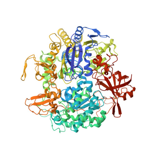
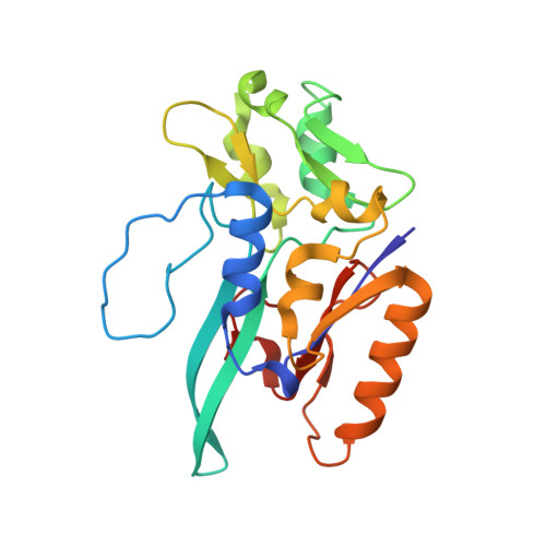
Gene Sequence and the 1.8 A Crystal Structure of the Tungsten-Containing Formate Dehydrogenase from Desulfovibrio Gigas

Raaijmakers, H.C.A.Macieira, S.Dias, J.Teixeira, S.Bursakov, S.Huber, R.Moura, J.Moura, I.Romao, M.

(2002) Structure 10: 1261

  • DOI: https://doi.org/10.1016/s0969-2126(02)00826-2
  • Primary Citation Related Structures: 
    1H0H

  • PubMed Abstract: 

    Desulfovibrio gigas formate dehydrogenase is the first representative of a tungsten-containing enzyme from a mesophile that has been structurally characterized. It is a heterodimer of 110 and 24 kDa subunits. The large subunit, homologous to E. coli FDH-H and to D. desulfuricans nitrate reductase, harbors the W site and one [4Fe-4S] center. No small subunit ortholog containing three [4Fe-4S] clusters has been reported. The structural homology with E. coli FDH-H shows that the essential residues (SeCys158, His159, and Arg407) at the active site are conserved. The active site is accessible via a positively charged tunnel, while product release may be facilitated, for H(+) by buried waters and protonable amino acids and for CO(2) through a hydrophobic channel.


  • Organizational Affiliation
    • REQUIMTE/CQFB, Departamento de Química, FCT, Universidade Nova de Lisboa, 2829-516 Caparica, Portugal.

Macromolecule Content 

  • Total Structure Weight: 274.27 kDa 
  • Atom Count: 21,055 
  • Modeled Residue Count: 2,382 
  • Deposited Residue Count: 2,382 
  • Unique protein chains: 2

Macromolecules

Find similar proteins by:|  3D Structure
Entity ID: 1
MoleculeChains  Sequence LengthOrganismDetailsImage
FORMATE DEHYDROGENASE SUBUNIT ALPHAA,
C [auth K]
977Megalodesulfovibrio gigasMutation(s): 0 
EC: 1.2.1.2 (PDB Primary Data), 1.17.1.9 (UniProt)
UniProt
Find proteins for Q934F5 (Megalodesulfovibrio gigas)
Explore Q934F5 
Go to UniProtKB:  Q934F5
Entity Groups
Sequence Clusters30% Identity50% Identity70% Identity90% Identity95% Identity100% Identity
UniProt GroupQ934F5
Sequence Annotations
Expand
Reference Sequence
Find similar proteins by:|  3D Structure
Entity ID: 2
MoleculeChains  Sequence LengthOrganismDetailsImage
FORMATE DEHYDROGENASE SUBUNIT BETAB,
D [auth L]
214Megalodesulfovibrio gigasMutation(s): 0 
UniProt
Find proteins for Q8GC87 (Megalodesulfovibrio gigas (strain ATCC 19364 / DSM 1382 / NCIMB 9332 / VKM B-1759))
Explore Q8GC87 
Go to UniProtKB:  Q8GC87
Entity Groups
Sequence Clusters30% Identity50% Identity70% Identity90% Identity95% Identity100% Identity
UniProt GroupQ8GC87
Sequence Annotations
Expand
Reference Sequence

Small Molecules

Ligands 7 Unique
IDChains Name / Formula / InChI Key2D Diagram3D Interactions
2MD

Query on 2MD


Download:Ideal Coordinates CCD File
F [auth A],
P [auth K]
GUANYLATE-O'-PHOSPHORIC ACID MONO-(2-AMINO-5,6-DIMERCAPTO-4-OXO-3,5,6,7,8A,9,10,10A-OCTAHYDRO-4H-8-OXA-1,3,9,10-TETRAAZA-ANTHRACEN-7-YLMETHYL) ESTER
C20 H28 N10 O13 P2 S2
YDYCAANTTOPADF-JKOCJXFLSA-N
MGD

Query on MGD


Download:Ideal Coordinates CCD File
G [auth A],
Q [auth K]
2-AMINO-5,6-DIMERCAPTO-7-METHYL-3,7,8A,9-TETRAHYDRO-8-OXA-1,3,9,10-TETRAAZA-ANTHRACEN-4-ONE GUANOSINE DINUCLEOTIDE
C20 H26 N10 O13 P2 S2
VQAGYJCYOLHZDH-ILXWUORBSA-N
SF4

Query on SF4


Download:Ideal Coordinates CCD File
I [auth A]
L [auth B]
M [auth B]
N [auth B]
S [auth K]
I [auth A],
L [auth B],
M [auth B],
N [auth B],
S [auth K],
V [auth L],
W [auth L],
X [auth L]
IRON/SULFUR CLUSTER
Fe4 S4
LJBDFODJNLIPKO-UHFFFAOYSA-N
EPE

Query on EPE


Download:Ideal Coordinates CCD File
K [auth A],
U [auth K]
4-(2-HYDROXYETHYL)-1-PIPERAZINE ETHANESULFONIC ACID
C8 H18 N2 O4 S
JKMHFZQWWAIEOD-UHFFFAOYSA-N
W

Query on W


Download:Ideal Coordinates CCD File
E [auth A],
O [auth K]
TUNGSTEN ION
W
FZFRVZDLZISPFJ-UHFFFAOYSA-N
CA

Query on CA


Download:Ideal Coordinates CCD File
J [auth A],
T [auth K]
CALCIUM ION
Ca
BHPQYMZQTOCNFJ-UHFFFAOYSA-N
UNX

Query on UNX


Download:Ideal Coordinates CCD File
H [auth A],
R [auth K]
UNKNOWN ATOM OR ION
X

Experimental Data & Validation

Experimental Data

  • Method: X-RAY DIFFRACTION
  • Resolution: 1.80 Å
  • R-Value Free:  0.201 (Depositor) 
  • R-Value Work:  0.167 (Depositor) 
  • R-Value Observed: 0.169 (Depositor) 
Space Group: P 1 21 1
Unit Cell:
Length ( Å )Angle ( ˚ )
a = 73.408α = 90
b = 110.398β = 93.53
c = 156.185γ = 90
Software Package:
Software NamePurpose
Omodel building
SCALEPACKdata scaling
SHARPphasing
AMoREphasing
ARP/wARPphasing
Ophasing
REFMACrefinement

Structure Validation

View Full Validation Report


Entry History 

Deposition Data

Revision History  (Full details and data files)

  • Version 1.0: 2003-02-20
    Type: Initial release
  • Version 1.1: 2014-01-29
    Changes: Atomic model, Database references, Derived calculations, Non-polymer description, Other, Structure summary, Version format compliance
  • Version 1.2: 2017-06-28
    Changes: Refinement description
  • Version 2.0: 2019-01-23
    Changes: Advisory, Atomic model, Data collection, Derived calculations
  • Version 2.1: 2019-04-03
    Changes: Data collection, Experimental preparation
  • Version 2.2: 2019-05-08
    Changes: Data collection, Experimental preparation
  • Version 2.3: 2023-12-13
    Changes: Advisory, Data collection, Database references, Derived calculations, Other, Refinement description
  • Version 2.4: 2024-10-23
    Changes: Structure summary