1H05 | pdb_00001h05

3-DEHYDROQUINATE DEHYDRATASE FROM MYCOBACTERIUM TUBERCULOSIS IN COMPLEX WITH SULPHATE


Experimental Data Snapshot

  • Method: X-RAY DIFFRACTION
  • Resolution: 1.50 Å
  • R-Value Free: 
    0.191 (Depositor) 
  • R-Value Work: 
    0.139 (Depositor), 0.149 (DCC) 

Starting Model: experimental
View more details

wwPDB Validation 3D Report Full Report

Validation slider image for 1H05

This is version 1.3 of the entry. See complete history

Literature

Specificity of Substrate Recognition by Type II Dehydroquinases as Revealed by Binding of Polyanions(1)

Evans, L.Roszak, A.W.Noble, L.Robinson, D.Chalk, P.Matthews, J.Coggins, J.R.Price, N.Lapthorn, A.J.

(2002) FEBS Lett 530: 24

  • DOI: https://doi.org/10.1016/s0014-5793(02)03346-x
  • Primary Citation Related Structures: 
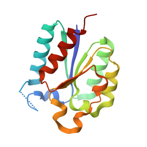
    1H05

  • PubMed Abstract: 

    The interactions between the polyanionic ligands phosphate and sulphate and the type II dehydroquinases from Streptomyces coelicolor and Mycobacterium tuberculosis have been characterised using a combination of structural and kinetic methods. From both approaches, it is clear that interactions are more complex in the case of the latter enzyme. The data provide new insights into the differences between the two enzymes in terms of substrate recognition and catalytic efficiency and may also explain the relative potencies of rationally designed inhibitors. An improved route to the synthesis of the substrate 3-dehydroquinic acid (dehydroquinate) is described.


  • Organizational Affiliation
    • Division of Biochemistry and Molecular Biology, Institute of Biomedical and Life Sciences, University of Glasgow, UK.

Macromolecule Content 

  • Total Structure Weight: 16.06 kDa 
  • Atom Count: 1,293 
  • Modeled Residue Count: 138 
  • Deposited Residue Count: 146 
  • Unique protein chains: 1

Macromolecules

Find similar proteins by:|  3D Structure
Entity ID: 1
MoleculeChains  Sequence LengthOrganismDetailsImage
3-DEHYDROQUINATE DEHYDRATASE146Mycobacterium tuberculosis H37RvMutation(s): 0 
EC: 4.2.1.10
UniProt
Find proteins for P9WPX7 (Mycobacterium tuberculosis (strain ATCC 25618 / H37Rv))
Explore P9WPX7 
Go to UniProtKB:  P9WPX7
Entity Groups
Sequence Clusters30% Identity50% Identity70% Identity90% Identity95% Identity100% Identity
UniProt GroupP9WPX7
Sequence Annotations
Expand
Reference Sequence

Experimental Data & Validation

Experimental Data

  • Method: X-RAY DIFFRACTION
  • Resolution: 1.50 Å
  • R-Value Free:  0.191 (Depositor) 
  • R-Value Work:  0.139 (Depositor), 0.149 (DCC) 
Space Group: F 2 3
Unit Cell:
Length ( Å )Angle ( ˚ )
a = 126.635α = 90
b = 126.635β = 90
c = 126.635γ = 90
Software Package:
Software NamePurpose
REFMACrefinement
DENZOdata reduction
SCALEPACKdata scaling
AMoREphasing

Structure Validation

View Full Validation Report


Entry History 

Deposition Data

Revision History  (Full details and data files)

  • Version 1.0: 2002-10-24
    Type: Initial release
  • Version 1.1: 2011-05-08
    Changes: Version format compliance
  • Version 1.2: 2011-07-13
    Changes: Version format compliance
  • Version 1.3: 2023-12-13
    Changes: Data collection, Database references, Derived calculations, Refinement description