1GYT | pdb_00001gyt

E. coli Aminopeptidase A (PepA)


Experimental Data Snapshot

  • Method: X-RAY DIFFRACTION
  • Resolution: 2.50 Å
  • R-Value Free: 
    0.209 (Depositor), 0.208 (DCC) 
  • R-Value Work: 
    0.165 (Depositor), 0.165 (DCC) 
  • R-Value Observed: 
    0.165 (Depositor) 

wwPDB Validation 3D Report Full Report

Validation slider image for 1GYT

This is version 1.6 of the entry. See complete history

Literature

X-Ray Structure of Aminopeptidase a from Escherichia Coli and a Model for the Nucleoprotein Complex in Xer Site-Specific Recombination

Straeter, N.Sherratt, D.J.Colloms, S.D.

(1999) EMBO J 18: 4513

  • DOI: https://doi.org/10.1093/emboj/18.16.4513
  • Primary Citation Related Structures: 
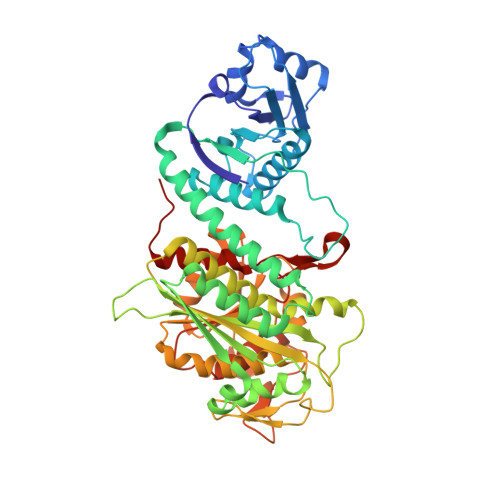
    1GYT

  • PubMed Abstract: 

    The structure of aminopeptidase A (PepA), which functions as a DNA-binding protein in Xer site-specific recombination and in transcriptional control of the carAB operon in Escherichia coli, has been determined at 2.5 A resolution. In Xer recombination at cer, PepA and the arginine repressor (ArgR) serve as accessory proteins, ensuring that recombination is exclusively intramolecular. In contrast, PepA homologues from other species have no known DNA-binding activity and are not implicated in transcriptional regulation or control of site-specific recombination. PepA comprises two domains, which have similar folds to the two domains of bovine lens leucine aminopeptidase (LAP). However, the N-terminal domain of PepA, which probably plays a significant role in DNA binding, is rotated by 19 degrees compared with its position in LAP. PepA is a homohexamer of 32 symmetry. A groove that runs from one trimer face across the 2-fold molecular axis to the other trimer face is proposed to be the DNA-binding site. Molecular modelling supports a structure of the Xer complex in which PepA, ArgR and a second PepA molecule are sandwiched along their 3-fold molecular axes, and the accessory sequences of the two recombination sites wrap around the accessory proteins as a right-handed superhelix such that three negative supercoils are trapped.


  • Organizational Affiliation
    • Institut für Kristallographie, Freie Universität Berlin, Takustrasse 6, 14195 Berlin, Germany. strater@chemie.fu-berlin.de

Macromolecule Content 

  • Total Structure Weight: 661.78 kDa 
  • Atom Count: 49,952 
  • Modeled Residue Count: 6,036 
  • Deposited Residue Count: 6,036 
  • Unique protein chains: 1

Macromolecules

Find similar proteins by:|  3D Structure
Entity ID: 1
MoleculeChains  Sequence LengthOrganismDetailsImage
CYTOSOL AMINOPEPTIDASE
A, B, C, D, E
A, B, C, D, E, F, G, H, I, J, K, L
503Escherichia coli K-12Mutation(s): 0 
EC: 3.4.11.1 (PDB Primary Data), 3.4.11.10 (UniProt)
UniProt
Find proteins for P68767 (Escherichia coli (strain K12))
Explore P68767 
Go to UniProtKB:  P68767
Entity Groups
Sequence Clusters30% Identity50% Identity70% Identity90% Identity95% Identity100% Identity
UniProt GroupP68767
Sequence Annotations
Expand
Reference Sequence

Small Molecules

Ligands 3 Unique
IDChains Name / Formula / InChI Key2D Diagram3D Interactions
ZN

Query on ZN


Download:Ideal Coordinates CCD File
AA [auth E]
CA [auth F]
DA [auth F]
GA [auth G]
HA [auth G]
AA [auth E],
CA [auth F],
DA [auth F],
GA [auth G],
HA [auth G],
KA [auth H],
LA [auth H],
M [auth A],
N [auth A],
NA [auth I],
OA [auth I],
P [auth B],
Q [auth B],
QA [auth J],
RA [auth J],
T [auth C],
TA [auth K],
U [auth C],
UA [auth K],
W [auth D],
X [auth D],
XA [auth L],
YA [auth L],
Z [auth E]
ZINC ION
Zn
PTFCDOFLOPIGGS-UHFFFAOYSA-N
CO3

Query on CO3


Download:Ideal Coordinates CCD File
BA [auth E]
EA [auth F]
IA [auth G]
MA [auth H]
O [auth A]
BA [auth E],
EA [auth F],
IA [auth G],
MA [auth H],
O [auth A],
PA [auth I],
R [auth B],
SA [auth J],
V [auth C],
VA [auth K],
Y [auth D],
ZA [auth L]
CARBONATE ION
C O3
BVKZGUZCCUSVTD-UHFFFAOYSA-L
CL

Query on CL


Download:Ideal Coordinates CCD File
FA [auth F],
JA [auth G],
S [auth B],
WA [auth K]
CHLORIDE ION
Cl
VEXZGXHMUGYJMC-UHFFFAOYSA-M

Experimental Data & Validation

Experimental Data

  • Method: X-RAY DIFFRACTION
  • Resolution: 2.50 Å
  • R-Value Free:  0.209 (Depositor), 0.208 (DCC) 
  • R-Value Work:  0.165 (Depositor), 0.165 (DCC) 
  • R-Value Observed: 0.165 (Depositor) 
Space Group: P 32
Unit Cell:
Length ( Å )Angle ( ˚ )
a = 178α = 90
b = 178β = 90
c = 244.4γ = 120
Software Package:
Software NamePurpose
CNSrefinement
DENZOdata reduction
SCALEPACKdata scaling

Structure Validation

View Full Validation Report


Entry History 

Deposition Data

  • Released Date: 2002-06-06 
  • Deposition Author(s): Straeter, N.

Revision History  (Full details and data files)

  • Version 1.0: 2002-06-06
    Type: Initial release
  • Version 1.1: 2011-05-08
    Changes: Version format compliance
  • Version 1.2: 2011-07-13
    Changes: Version format compliance
  • Version 1.3: 2017-07-12
    Changes: Derived calculations
  • Version 1.4: 2018-02-07
    Changes: Source and taxonomy
  • Version 1.5: 2019-07-24
    Changes: Data collection
  • Version 1.6: 2024-05-08
    Changes: Data collection, Database references, Derived calculations, Other