1GY8 | pdb_00001gy8

Trypanosoma brucei UDP-galactose 4' epimerase


Experimental Data Snapshot

  • Method: X-RAY DIFFRACTION
  • Resolution: 2.00 Å
  • R-Value Free: 
    0.245 (Depositor) 
  • R-Value Work: 
    0.216 (Depositor), 0.229 (DCC) 
  • R-Value Observed: 
    0.216 (Depositor) 

Starting Model: experimental
View more details

wwPDB Validation 3D Report Full Report

Validation slider image for 1GY8

Ligand Structure Quality Assessment 


This is version 1.3 of the entry. See complete history

Literature

High-Resolution Crystal Structure of Trypanosoma Brucei Udp-Galactose 4'-Epimerase: A Potential Target for Structure-Based Development of Novel Trypanocides

Shaw, M.P.Bond, C.S.Roper, J.R.Gourley, D.G.Ferguson, M.A.J.Hunter, W.N.

(2003) Mol Biochem Parasitol 126: 173

  • DOI: https://doi.org/10.1016/s0166-6851(02)00243-8
  • Primary Citation Related Structures: 
    1GY8

  • PubMed Abstract: 

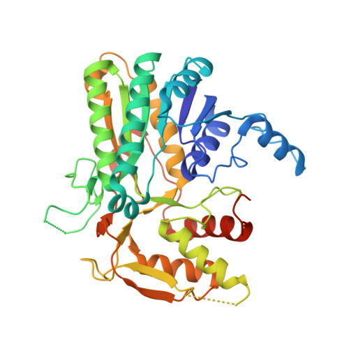
    The crystal structure of UDP-galactose 4'-epimerase from the protozoan parasite Trypanosoma brucei in complex with the cofactor NAD(+) and a fragment of the substrates, UDP, has been determined at 2.0 A resolution (1 A = 0.1 nm). This enzyme, recently proven to be essential for this pathogenic parasite, shares 33% sequence identity with the corresponding enzyme in the human host. Structural comparisons indicate that many of the protein-ligand interactions are conserved between the two enzymes. However, in the UDP-binding pocket there is a non-conservative substitution from Gly237 in the human enzyme to Cys266 in the T. brucei enzyme. Such a significant difference could be exploited by the structure-based design of selective inhibitors using the structure of the trypanosomatid enzyme as a template.


  • Organizational Affiliation
    • Division of Biological Chemistry and Molecular Microbiology, School of Life Sciences, University of Dundee, Dundee DD1 5EH, UK.

Macromolecule Content 

  • Total Structure Weight: 179.5 kDa 
  • Atom Count: 12,972 
  • Modeled Residue Count: 1,467 
  • Deposited Residue Count: 1,588 
  • Unique protein chains: 1

Macromolecules

Find similar proteins by:|  3D Structure
Entity ID: 1
MoleculeChains  Sequence LengthOrganismDetailsImage
UDP-GALACTOSE 4-EPIMERASE
A, B, C, D
397Trypanosoma bruceiMutation(s): 0 
EC: 5.1.3.2
UniProt
Find proteins for Q8T8E9 (Trypanosoma brucei)
Explore Q8T8E9 
Go to UniProtKB:  Q8T8E9
Entity Groups
Sequence Clusters30% Identity50% Identity70% Identity90% Identity95% Identity100% Identity
UniProt GroupQ8T8E9
Sequence Annotations
Expand
Reference Sequence

Small Molecules

Ligands 2 Unique
IDChains Name / Formula / InChI Key2D Diagram3D Interactions
NAD

Query on NAD


Download:Ideal Coordinates CCD File
E [auth A],
G [auth B],
I [auth C],
K [auth D]
NICOTINAMIDE-ADENINE-DINUCLEOTIDE
C21 H27 N7 O14 P2
BAWFJGJZGIEFAR-NNYOXOHSSA-N
UDP

Query on UDP


Download:Ideal Coordinates CCD File
F [auth A],
H [auth B],
J [auth C],
L [auth D]
URIDINE-5'-DIPHOSPHATE
C9 H14 N2 O12 P2
XCCTYIAWTASOJW-XVFCMESISA-N

Experimental Data & Validation

Experimental Data

  • Method: X-RAY DIFFRACTION
  • Resolution: 2.00 Å
  • R-Value Free:  0.245 (Depositor) 
  • R-Value Work:  0.216 (Depositor), 0.229 (DCC) 
  • R-Value Observed: 0.216 (Depositor) 
Space Group: P 21 21 21
Unit Cell:
Length ( Å )Angle ( ˚ )
a = 102.185α = 90
b = 112.533β = 90
c = 160.591γ = 90
Software Package:
Software NamePurpose
CNSrefinement
DENZOdata reduction
SCALEPACKdata scaling
AMoREphasing

Structure Validation

View Full Validation Report


Ligand Structure Quality Assessment 


Entry History 

Deposition Data

Revision History  (Full details and data files)

  • Version 1.0: 2003-05-22
    Type: Initial release
  • Version 1.1: 2011-05-08
    Changes: Version format compliance
  • Version 1.2: 2011-07-13
    Changes: Version format compliance
  • Version 1.3: 2023-12-13
    Changes: Data collection, Database references, Other, Refinement description