1GX4 | pdb_00001gx4

ALPHA-,1,3 GALACTOSYLTRANSFERASE - N-ACETYL LACTOSAMINE COMPLEX


Experimental Data Snapshot

  • Method: X-RAY DIFFRACTION
  • Resolution: 1.46 Å
  • R-Value Free: 
    0.194 (Depositor) 
  • R-Value Work: 
    0.138 (DCC) 
  • R-Value Observed: 
    0.149 (Depositor) 

wwPDB Validation 3D Report Full Report

Validation slider image for 1GX4

Ligand Structure Quality Assessment 


This is version 2.1 of the entry. See complete history

Literature

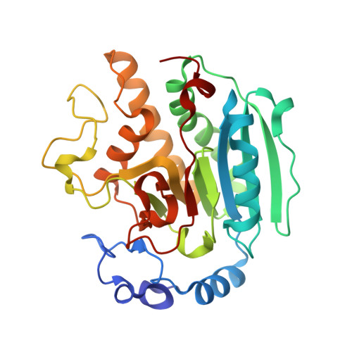
Structural Basis of Ordered Binding of Donor and Acceptor Substrates to the Retaining Glycosyltransferase, Alpha -1,3 Galactosyltransferase

Boix, E.Zhang, Y.Swaminathan, G.J.Brew, K.Acharya, K.R.

(2002) J Biological Chem 277: 28310

  • DOI: https://doi.org/10.1074/jbc.M202631200
  • Primary Citation Related Structures: 
    1GWV, 1GWW, 1GX0, 1GX4

  • PubMed Abstract: 

    Bovine alpha-1,3-galactosyltransferase (alpha3GT) catalyzes the synthesis of the alpha-galactose (alpha-Gal) epitope, the target of natural human antibodies. It represents a family of enzymes, including the histo blood group A and B transferases, that catalyze retaining glycosyltransfer reactions of unknown mechanism. An initial study of alpha3GT in a crystal form with limited resolution and considerable disorder suggested the possible formation of a beta-galactosyl-enzyme covalent intermediate (Gastinel, L. N., Bignon, C., Misra, A. K., Hindsgaul, O., Shaper, J. H., and Joziasse, D. H. (2001) EMBO J. 20, 638-649). Highly ordered structures are described for complexes of alpha3GT with donor substrate, UDP-galactose, UDP- glucose, and two acceptor substrates, lactose and N-acetyllactosamine, at resolutions up to 1.46 A. Structural and calorimetric binding studies suggest an obligatory ordered binding of donor and acceptor substrates, linked to a donor substrate-induced conformational change, and the direct participation of UDP in acceptor binding. The monosaccharide-UDP bond is cleaved in the structures containing UDP-galactose and UDP-glucose, producing non-covalent complexes containing buried beta-galactose and alpha-glucose. The location of these monosaccharides and molecular modeling suggest that binding of a distorted conformation of UDP-galactose may be important in the catalytic mechanism of alpha3GT.


  • Organizational Affiliation
    • Department of Biology and Biochemistry, University of Bath, Claverton Down, Bath BA2 7AY, United Kingdom.

Macromolecule Content 

  • Total Structure Weight: 70.23 kDa 
  • Atom Count: 5,763 
  • Modeled Residue Count: 574 
  • Deposited Residue Count: 578 
  • Unique protein chains: 1

Macromolecules

Find similar proteins by:|  3D Structure
Entity ID: 1
MoleculeChains  Sequence LengthOrganismDetailsImage
N-ACETYLLACTOSAMINIDE ALPHA-1,3-GALACTOSYLTRANSFERASE
A, B
289Bos taurusMutation(s): 0 
EC: 2.4.1.151 (PDB Primary Data), 2.4.1.87 (UniProt)
UniProt
Find proteins for P14769 (Bos taurus)
Explore P14769 
Go to UniProtKB:  P14769
Entity Groups
Sequence Clusters30% Identity50% Identity70% Identity90% Identity95% Identity100% Identity
UniProt GroupP14769
Sequence Annotations
Expand
Reference Sequence

Oligosaccharides

Help  
Entity ID: 2
MoleculeChains Length2D Diagram GlycosylationD Interactions
beta-D-galactopyranose-(1-4)-2-acetamido-2-deoxy-beta-D-glucopyranose
C, D
2N/A
Glycosylation Resources
GlyTouCan: G00055MO
GlyCosmos: G00055MO
GlyGen: G00055MO

Small Molecules

Ligands 3 Unique
IDChains Name / Formula / InChI Key2D Diagram3D Interactions
UDP

Query on UDP


Download:Ideal Coordinates CCD File
E [auth A],
J [auth B]
URIDINE-5'-DIPHOSPHATE
C9 H14 N2 O12 P2
XCCTYIAWTASOJW-XVFCMESISA-N
GOL

Query on GOL


Download:Ideal Coordinates CCD File
G [auth A],
H [auth A],
I [auth A],
L [auth B]
GLYCEROL
C3 H8 O3
PEDCQBHIVMGVHV-UHFFFAOYSA-N
MN

Query on MN


Download:Ideal Coordinates CCD File
F [auth A],
K [auth B]
MANGANESE (II) ION
Mn
WAEMQWOKJMHJLA-UHFFFAOYSA-N

Experimental Data & Validation

Experimental Data

  • Method: X-RAY DIFFRACTION
  • Resolution: 1.46 Å
  • R-Value Free:  0.194 (Depositor) 
  • R-Value Work:  0.138 (DCC) 
  • R-Value Observed: 0.149 (Depositor) 
Space Group: P 1 21 1
Unit Cell:
Length ( Å )Angle ( ˚ )
a = 45.626α = 90
b = 95.245β = 99.09
c = 95.638γ = 90
Software Package:
Software NamePurpose
SHELXL-97refinement
DENZOdata reduction
SCALEPACKdata scaling
AMoREphasing

Structure Validation

View Full Validation Report


Ligand Structure Quality Assessment 


Entry History 

Deposition Data

Revision History  (Full details and data files)

  • Version 1.0: 2003-03-27
    Type: Initial release
  • Version 1.1: 2011-09-07
    Changes: Atomic model, Database references, Derived calculations, Non-polymer description, Other, Source and taxonomy, Structure summary, Version format compliance
  • Version 1.2: 2019-05-22
    Changes: Data collection, Other, Refinement description
  • Version 2.0: 2020-07-29
    Type: Remediation
    Reason: Carbohydrate remediation
    Changes: Atomic model, Data collection, Derived calculations, Structure summary
  • Version 2.1: 2024-05-08
    Changes: Data collection, Database references, Structure summary