1GUE | pdb_00001gue

SENSORY RHODOPSIN II


Experimental Data Snapshot

  • Method: X-RAY DIFFRACTION
  • Resolution: 2.27 Å
  • R-Value Free: 
    0.311 (Depositor) 
  • R-Value Work: 
    0.277 (Depositor) 
  • R-Value Observed: 
    0.277 (Depositor) 

Starting Model: experimental
View more details

wwPDB Validation 3D Report Full Report

Validation slider image for 1GUE

This is version 1.4 of the entry. See complete history

Literature

Early Structural Rearrangements in the Photocycle of an Integral Membrane Sensory Receptor

Edman, K.Royant, A.Nollert, P.Maxwell, C.A.Pebay-Peyroula, E.Navarro, J.Neutze, R.Landau, E.M.

(2002) Structure 10: 473

  • DOI: https://doi.org/10.1016/s0969-2126(02)00736-0
  • Primary Citation Related Structures: 
    1GU8, 1GUE

  • PubMed Abstract: 

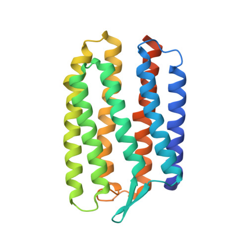
    Sensory rhodopsins are the primary receptors of vision in animals and phototaxis in microorganisms. Light triggers the rapid isomerization of a buried retinal chromophore, which the protein both accommodates and amplifies into the larger structural rearrangements required for signaling. We trapped an early intermediate of the photocycle of sensory rhodopsin II from Natronobacterium pharaonis (pSRII) in 3D crystals and determined its X-ray structure to 2.3 A resolution. The observed structural rearrangements were localized near the retinal chromophore, with a key water molecule becoming disordered and the retinal's beta-ionone ring undergoing a prominent movement. Comparison with the early structural rearrangements of bacteriorhodopsin illustrates how modifications in the retinal binding pocket of pSRII allow subtle differences in the early relaxation of photoisomerized retinal.


  • Organizational Affiliation
    • Department of Molecular Biotechnology, Chalmers University of Technology, Box 462, S-40530 Gothenburg, Sweden.

Macromolecule Content 

  • Total Structure Weight: 25.69 kDa 
  • Atom Count: 1,660 
  • Modeled Residue Count: 218 
  • Deposited Residue Count: 239 
  • Unique protein chains: 1

Macromolecules

Find similar proteins by:|  3D Structure
Entity ID: 1
MoleculeChains  Sequence LengthOrganismDetailsImage
SENSORY RHODOPSIN II239Natronomonas pharaonisMutation(s): 0 
Membrane Entity: Yes 
UniProt
Find proteins for P42196 (Natronomonas pharaonis)
Explore P42196 
Go to UniProtKB:  P42196
Entity Groups
Sequence Clusters30% Identity50% Identity70% Identity90% Identity95% Identity100% Identity
UniProt GroupP42196
Sequence Annotations
Expand
Reference Sequence

Small Molecules

Ligands 2 Unique
IDChains Name / Formula / InChI Key2D Diagram3D Interactions
RET

Query on RET


Download:Ideal Coordinates CCD File
B [auth A]RETINAL
C20 H28 O
NCYCYZXNIZJOKI-OVSJKPMPSA-N
CL

Query on CL


Download:Ideal Coordinates CCD File
C [auth A]CHLORIDE ION
Cl
VEXZGXHMUGYJMC-UHFFFAOYSA-M

Experimental Data & Validation

Experimental Data

  • Method: X-RAY DIFFRACTION
  • Resolution: 2.27 Å
  • R-Value Free:  0.311 (Depositor) 
  • R-Value Work:  0.277 (Depositor) 
  • R-Value Observed: 0.277 (Depositor) 
Space Group: C 2 2 21
Unit Cell:
Length ( Å )Angle ( ˚ )
a = 85.51α = 90
b = 128.553β = 90
c = 50.799γ = 90
Software Package:
Software NamePurpose
CNSrefinement
DENZOdata reduction
CCP4data scaling
AMoREphasing

Structure Validation

View Full Validation Report


Entry History 

Deposition Data

Revision History  (Full details and data files)

  • Version 1.0: 2002-04-12
    Type: Initial release
  • Version 1.1: 2011-05-08
    Changes: Version format compliance
  • Version 1.2: 2011-07-13
    Changes: Version format compliance
  • Version 1.3: 2023-12-13
    Changes: Data collection, Database references, Derived calculations, Refinement description
  • Version 1.4: 2024-11-20
    Changes: Structure summary