1GU1 | pdb_00001gu1

Crystal structure of type II dehydroquinase from Streptomyces coelicolor complexed with 2,3-anhydro-quinic acid


Experimental Data Snapshot

  • Method: X-RAY DIFFRACTION
  • Resolution: 1.80 Å
  • R-Value Free: 
    0.200 (Depositor) 
  • R-Value Work: 
    0.155 (Depositor) 

Starting Model: experimental
View more details

wwPDB Validation 3D Report Full Report

Validation slider image for 1GU1

This is version 1.3 of the entry. See complete history

Literature

The Structure and Mechanism of the Type II Dehydroquinase from Streptomyces Coelicolor

Roszak, A.W.Robinson, D.A.Krell, T.Hunter, I.S.Fredrickson, M.Abell, C.Coggins, J.R.Lapthorn, A.J.

(2002) Structure 10: 493

  • DOI: https://doi.org/10.1016/s0969-2126(02)00747-5
  • Primary Citation Related Structures: 
    1D0I, 1GTZ, 1GU0, 1GU1

  • PubMed Abstract: 

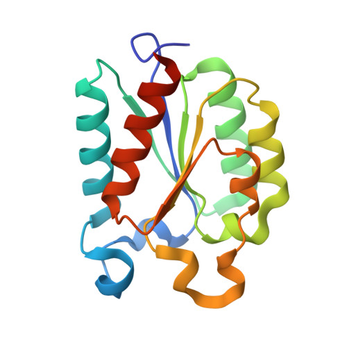
    The structure of the type II DHQase from Streptomyces coelicolor has been solved and refined to high resolution in complexes with a number of ligands, including dehydroshikimate and a rationally designed transition state analogue, 2,3-anhydro-quinic acid. These structures define the active site of the enzyme and the role of key amino acid residues and provide snap shots of the catalytic cycle. The resolution of the flexible lid domain (residues 21-31) shows that the invariant residues Arg23 and Tyr28 close over the active site cleft. The tyrosine acts as the base in the initial proton abstraction, and evidence is provided that the reaction proceeds via an enol intermediate. The active site of the structure of DHQase in complex with the transition state analog also includes molecules of tartrate and glycerol, which provide a basis for further inhibitor design.


  • Organizational Affiliation
    • Department of Chemistry, Institute of Biomedical and Life Sciences, University of Glasgow, Scotland, United Kingdom.

Macromolecule Content 

  • Total Structure Weight: 204.32 kDa 
  • Atom Count: 16,578 
  • Modeled Residue Count: 1,788 
  • Deposited Residue Count: 1,872 
  • Unique protein chains: 1

Macromolecules

Find similar proteins by:|  3D Structure
Entity ID: 1
MoleculeChains  Sequence LengthOrganismDetailsImage
3-DEHYDROQUINATE DEHYDRATASE
A, B, C, D, E
A, B, C, D, E, F, G, H, I, J, K, L
156Streptomyces coelicolorMutation(s): 0 
EC: 4.2.1.10
UniProt
Find proteins for P15474 (Streptomyces coelicolor (strain ATCC BAA-471 / A3(2) / M145))
Explore P15474 
Go to UniProtKB:  P15474
Entity Groups
Sequence Clusters30% Identity50% Identity70% Identity90% Identity95% Identity100% Identity
UniProt GroupP15474
Sequence Annotations
Expand
Reference Sequence

Small Molecules

Ligands 4 Unique
IDChains Name / Formula / InChI Key2D Diagram3D Interactions
FA1

Query on FA1


Download:Ideal Coordinates CCD File
AA [auth E]
DA [auth F]
GA [auth G]
JA [auth H]
M [auth A]
AA [auth E],
DA [auth F],
GA [auth G],
JA [auth H],
M [auth A],
NA [auth I],
Q [auth B],
QA [auth J],
TA [auth K],
U [auth C],
WA [auth L],
X [auth D]
2,3 -ANHYDRO-QUINIC ACID
C7 H10 O5
VTEDVYGIJPLVFF-XAHCXIQSSA-N
TLA

Query on TLA


Download:Ideal Coordinates CCD File
CA [auth E]
FA [auth F]
IA [auth G]
LA [auth H]
O [auth A]
CA [auth E],
FA [auth F],
IA [auth G],
LA [auth H],
O [auth A],
PA [auth I],
S [auth B],
SA [auth J],
VA [auth K],
W [auth C],
YA [auth L],
Z [auth D]
L(+)-TARTARIC ACID
C4 H6 O6
FEWJPZIEWOKRBE-JCYAYHJZSA-N
TRS

Query on TRS


Download:Ideal Coordinates CCD File
MA [auth H],
P [auth A],
T [auth B],
ZA [auth L]
2-AMINO-2-HYDROXYMETHYL-PROPANE-1,3-DIOL
C4 H12 N O3
LENZDBCJOHFCAS-UHFFFAOYSA-O
GOL

Query on GOL


Download:Ideal Coordinates CCD File
BA [auth E]
EA [auth F]
HA [auth G]
KA [auth H]
N [auth A]
BA [auth E],
EA [auth F],
HA [auth G],
KA [auth H],
N [auth A],
OA [auth I],
R [auth B],
RA [auth J],
UA [auth K],
V [auth C],
XA [auth L],
Y [auth D]
GLYCEROL
C3 H8 O3
PEDCQBHIVMGVHV-UHFFFAOYSA-N

Experimental Data & Validation

Experimental Data

  • Method: X-RAY DIFFRACTION
  • Resolution: 1.80 Å
  • R-Value Free:  0.200 (Depositor) 
  • R-Value Work:  0.155 (Depositor) 
Space Group: P 21 21 21
Unit Cell:
Length ( Å )Angle ( ˚ )
a = 113.6α = 90
b = 138.4β = 90
c = 141.5γ = 90
Software Package:
Software NamePurpose
REFMACrefinement
DENZOdata reduction
SCALEPACKdata scaling
AMoREphasing

Structure Validation

View Full Validation Report


Entry History 

Deposition Data

Revision History  (Full details and data files)

  • Version 1.0: 2002-04-12
    Type: Initial release
  • Version 1.1: 2011-09-28
    Changes: Derived calculations, Non-polymer description, Other, Version format compliance
  • Version 1.2: 2013-07-24
    Changes: Data collection, Structure summary
  • Version 1.3: 2023-12-13
    Changes: Data collection, Database references, Derived calculations, Refinement description