1GT7 | pdb_00001gt7

L-rhamnulose-1-phosphate aldolase from Escherichia coli


Experimental Data Snapshot

  • Method: X-RAY DIFFRACTION
  • Resolution: 2.70 Å
  • R-Value Free: 
    0.235 (Depositor), 0.225 (DCC) 
  • R-Value Work: 
    0.233 (Depositor), 0.219 (DCC) 

wwPDB Validation 3D Report Full Report

Validation slider image for 1GT7

Ligand Structure Quality Assessment 


This is version 1.4 of the entry. See complete history

Literature

The Structure of L-Rhamnulose-1-Phosphate Aldolase (Class II) Solved by Low-Resolution Sir Phasing and 20-Fold Ncs Averaging

Kroemer, M.Schulz, G.E.

(2002) Acta Crystallogr D Biol Crystallogr 58: 824

  • DOI: https://doi.org/10.1107/s0907444902004614
  • Primary Citation Related Structures: 
    1GT7

  • PubMed Abstract: 

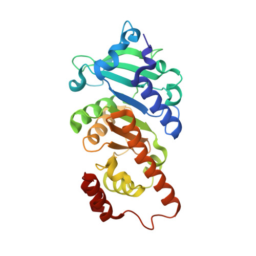
    The enzyme L-rhamnulose-1-phosphate aldolase catalyzes the reversible cleavage of L-rhamnulose-1-phosphate to dihydroxyacetone phosphate and L-lactaldehyde. It is a homotetramer with an M(r) of 30 000 per subunit and crystallized in space group P3(2)21. The enzyme shows a low sequence identity of 18% with the structurally known L-fuculose-1-phosphate aldolase that splits a stereoisomer in a similar reaction. Structure analysis was initiated with a single heavy-atom derivative measured to 6 A resolution. The resulting poor electron density, a self-rotation function and the working hypothesis that both enzymes are C(4) symmetric with envelopes that resemble one another allowed the location of the 20 protomers of the asymmetric unit. The crystal-packing unit was a D(4)-symmetric propeller consisting of five D(4)-symmetric octamers around an internal crystallographic twofold axis. Presumably, the propellers associate laterally in layers, which in turn pile up along the 3(2) axis to form the crystal. The non-crystallographic symmetry was used to extend the phases to the 2.7 A resolution limit and to establish a refined atomic model of the enzyme. The structure showed that the two enzymes are indeed homologous and that they possess chemically similar active centres.


  • Organizational Affiliation
    • Institut für Organische Chemie und Biochemie, Albert-Ludwigs-Universität Freiburg, Albertstrasse 21, 79104 Freiburg im Breisgau, Germany.

Macromolecule Content 

  • Total Structure Weight: 608.22 kDa 
  • Atom Count: 46,180 
  • Modeled Residue Count: 5,480 
  • Deposited Residue Count: 5,480 
  • Unique protein chains: 1

Macromolecules

Find similar proteins by:|  3D Structure
Entity ID: 1
MoleculeChains  Sequence LengthOrganismDetailsImage
RHAMNULOSE-1-PHOSPHATE ALDOLASE
A, B, C, D, E
A, B, C, D, E, F, G, H, I, J, K, L, M, N, O, P, Q, R, S, T
274Escherichia coliMutation(s): 0 
EC: 4.1.2.19
UniProt
Find proteins for P32169 (Escherichia coli (strain K12))
Explore P32169 
Go to UniProtKB:  P32169
Entity Groups
Sequence Clusters30% Identity50% Identity70% Identity90% Identity95% Identity100% Identity
UniProt GroupP32169
Sequence Annotations
Expand
Reference Sequence

Small Molecules

Ligands 2 Unique
IDChains Name / Formula / InChI Key2D Diagram3D Interactions
PGH

Query on PGH


Download:Ideal Coordinates CCD File
BA [auth D]
BB [auth Q]
DA [auth E]
DB [auth R]
FA [auth F]
BA [auth D],
BB [auth Q],
DA [auth E],
DB [auth R],
FA [auth F],
FB [auth S],
HA [auth G],
HB [auth T],
JA [auth H],
LA [auth I],
NA [auth J],
PA [auth K],
RA [auth L],
TA [auth M],
V [auth A],
VA [auth N],
X [auth B],
XA [auth O],
Z [auth C],
ZA [auth P]
PHOSPHOGLYCOLOHYDROXAMIC ACID
C2 H6 N O6 P
BAXHHWZKQZIJID-UHFFFAOYSA-N
ZN

Query on ZN


Download:Ideal Coordinates CCD File
AA [auth D]
AB [auth Q]
CA [auth E]
CB [auth R]
EA [auth F]
AA [auth D],
AB [auth Q],
CA [auth E],
CB [auth R],
EA [auth F],
EB [auth S],
GA [auth G],
GB [auth T],
IA [auth H],
KA [auth I],
MA [auth J],
OA [auth K],
QA [auth L],
SA [auth M],
U [auth A],
UA [auth N],
W [auth B],
WA [auth O],
Y [auth C],
YA [auth P]
ZINC ION
Zn
PTFCDOFLOPIGGS-UHFFFAOYSA-N

Experimental Data & Validation

Experimental Data

  • Method: X-RAY DIFFRACTION
  • Resolution: 2.70 Å
  • R-Value Free:  0.235 (Depositor), 0.225 (DCC) 
  • R-Value Work:  0.233 (Depositor), 0.219 (DCC) 
Space Group: P 32 2 1
Unit Cell:
Length ( Å )Angle ( ˚ )
a = 225.76α = 90
b = 225.76β = 90
c = 285.645γ = 120
Software Package:
Software NamePurpose
DMmodel building
SCALAdata scaling
SCALEITphasing
MLPHAREphasing
GETAXphasing
DMphasing
REFMACrefinement

Structure Validation

View Full Validation Report


Ligand Structure Quality Assessment 


Entry History 

Deposition Data

Revision History  (Full details and data files)

  • Version 1.0: 2002-05-03
    Type: Initial release
  • Version 1.1: 2013-11-20
    Changes: Derived calculations, Non-polymer description, Other, Version format compliance
  • Version 1.2: 2019-05-08
    Changes: Data collection, Experimental preparation, Other
  • Version 1.3: 2019-07-24
    Changes: Data collection
  • Version 1.4: 2024-05-08
    Changes: Data collection, Database references, Derived calculations, Other