1GRC | pdb_00001grc

CRYSTAL STRUCTURE OF GLYCINAMIDE RIBONUCLEOTIDE TRANSFORMYLASE FROM ESCHERICHIA COLI AT 3.0 ANGSTROMS RESOLUTION: A TARGET ENZYME FOR CHEMOTHERAPY


Experimental Data Snapshot

  • Method: X-RAY DIFFRACTION
  • Resolution: 3.00 Å
  • R-Value Work: 
    0.190 (Depositor) 
  • R-Value Observed: 
    0.190 (Depositor) 

wwPDB Validation 3D Report Full Report

Validation slider image for 1GRC

This is version 1.3 of the entry. See complete history

Literature

Crystal structure of glycinamide ribonucleotide transformylase from Escherichia coli at 3.0 A resolution. A target enzyme for chemotherapy.

Chen, P.Schulze-Gahmen, U.Stura, E.A.Inglese, J.Johnson, D.L.Marolewski, A.Benkovic, S.J.Wilson, I.A.

(1992) J Mol Biology 227: 283-292

  • DOI: https://doi.org/10.1016/0022-2836(92)90698-j
  • Primary Citation Related Structures: 
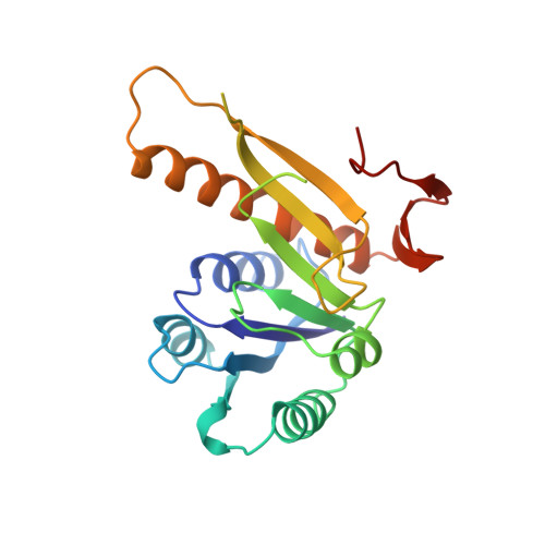
    1GRC

  • PubMed Abstract: 

    The atomic structure of glycinamide ribonucleotide transformylase, an essential enzyme in purine biosynthesis, has been determined at 3.0 A resolution. The last three C-terminal residues and a sequence stretch of 18 residues (residues 113 to 130) are not visible in the electron density map. The enzyme forms a dimer in the crystal structure. Each monomer is divided into two domains, which are connected by a central mainly parallel seven-stranded beta-sheet. The N-terminal domain contains a Rossmann type mononucleotide fold with a phosphate ion bound to the C-terminal end of the first beta-strand. A long narrow cleft stretches from the phosphate to a conserved aspartic acid, Asp144, which has been suggested as an active-site residue. The cleft is lined by a cluster of residues, which are conserved between bacterial, yeast, avian and human enzymes, and likely represents the binding pocket and active site of the enzyme. GAR Tfase binds a reduced folate cofactor and glycinamide ribonucleotide for the catalysis of one of the initial steps in purine biosynthesis. Folate analogs and multi-substrate inhibitors of the enzyme have antineoplastic effects and the structure determination of the unliganded enzyme and enzyme-inhibitor complexes will aid the development of anti-cancer drugs.


  • Organizational Affiliation
    • Department of Molecular Biology, Scripps Research Institute, La Jolla, CA 92037.

Macromolecule Content 

  • Total Structure Weight: 46.72 kDa 
  • Atom Count: 2,963 
  • Modeled Residue Count: 386 
  • Deposited Residue Count: 424 
  • Unique protein chains: 1

Macromolecules

Find similar proteins by:|  3D Structure
Entity ID: 1
MoleculeChains  Sequence LengthOrganismDetailsImage
GLYCINAMIDE RIBONUCLEOTIDE TRANSFORMYLASE
A, B
212Escherichia coliMutation(s): 0 
EC: 2.1.2.2
UniProt
Find proteins for P08179 (Escherichia coli (strain K12))
Explore P08179 
Go to UniProtKB:  P08179
Entity Groups
Sequence Clusters30% Identity50% Identity70% Identity90% Identity95% Identity100% Identity
UniProt GroupP08179
Sequence Annotations
Expand
Reference Sequence

Small Molecules

Experimental Data & Validation

Experimental Data

  • Method: X-RAY DIFFRACTION
  • Resolution: 3.00 Å
  • R-Value Work:  0.190 (Depositor) 
  • R-Value Observed: 0.190 (Depositor) 
Space Group: C 2 2 21
Unit Cell:
Length ( Å )Angle ( ˚ )
a = 140.5α = 90
b = 98.2β = 90
c = 103.3γ = 90
Software Package:
Software NamePurpose
X-PLORmodel building
X-PLORrefinement
X-PLORphasing

Structure Validation

View Full Validation Report


Entry History 

Deposition Data

Revision History  (Full details and data files)

  • Version 1.0: 1993-10-31
    Type: Initial release
  • Version 1.1: 2008-03-24
    Changes: Version format compliance
  • Version 1.2: 2011-07-13
    Changes: Version format compliance
  • Version 1.3: 2024-02-07
    Changes: Data collection, Database references, Derived calculations, Other