1GQT | pdb_00001gqt

Activation of Ribokinase by Monovalent Cations


Experimental Data Snapshot

  • Method: X-RAY DIFFRACTION
  • Resolution: 2.34 Å
  • R-Value Free: 
    0.257 (Depositor), 0.256 (DCC) 
  • R-Value Work: 
    0.193 (Depositor), 0.195 (DCC) 
  • R-Value Observed: 
    0.196 (Depositor) 

wwPDB Validation 3D Report Full Report

Validation slider image for 1GQT

Ligand Structure Quality Assessment 


This is version 3.1 of the entry. See complete history

Literature

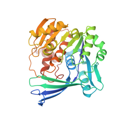
Activation of Ribokinase by Monovalent Cations.

Andersson, C.E.Mowbray, S.L.

(2002) J Mol Biology 315: 409

  • DOI: https://doi.org/10.1006/jmbi.2001.5248
  • Primary Citation Related Structures: 
    1GQT

  • PubMed Abstract: 

    Carbohydrate kinases frequently require a monovalent cation for their activity. The physical basis of this phenomenon is, however, usually unclear. We report here that Escherichia coli ribokinase is activated by potassium with an apparent K(d) of 5 mM; the enzyme should therefore be fully activated under physiological conditions. Cesium can be used as an alternative ion, with an apparent K(d) of 17 mM. An X-ray structure of ribokinase in the presence of cesium was solved and refined at 2.34 A resolution. The cesium ion was bound between two loops immediately adjacent to the anion hole of the active site. The buried location of the site suggests that conformational changes will accompany ion binding, thus providing a direct mechanism for activation. Comparison with structures of a related enzyme, the adenosine kinase of Toxoplasma gondii, support this proposal. This is apparently the first instance in which conformational activation of a carbohydrate kinase by a monovalent cation has been assigned a clear structural basis. The mechanism is probably general to ribokinases, to some adenosine kinases, and to other members of the larger family. A careful re-evaluation of the biochemical and structural data is suggested for other enzyme systems.


  • Organizational Affiliation
    • Department of Cell and Molecular Biology, Uppsala University, S-751 24 Uppsala, Sweden.

Macromolecule Content 

  • Total Structure Weight: 131.93 kDa 
  • Atom Count: 9,352 
  • Modeled Residue Count: 1,223 
  • Deposited Residue Count: 1,236 
  • Unique protein chains: 1

Macromolecules

Find similar proteins by:|  3D Structure
Entity ID: 1
MoleculeChains  Sequence LengthOrganismDetailsImage
RIBOKINASE
A, B, C, D
309Escherichia coli K-12Mutation(s): 0 
EC: 2.7.1.15
UniProt
Find proteins for P0A9J6 (Escherichia coli (strain K12))
Explore P0A9J6 
Go to UniProtKB:  P0A9J6
Entity Groups
Sequence Clusters30% Identity50% Identity70% Identity90% Identity95% Identity100% Identity
UniProt GroupP0A9J6
Sequence Annotations
Expand
Reference Sequence

Small Molecules

Ligands 3 Unique
IDChains Name / Formula / InChI Key2D Diagram3D Interactions
ACP

Query on ACP


Download:Ideal Coordinates CCD File
F [auth A],
I [auth B],
L [auth C]
PHOSPHOMETHYLPHOSPHONIC ACID ADENYLATE ESTER
C11 H18 N5 O12 P3
UFZTZBNSLXELAL-IOSLPCCCSA-N
RIB

Query on RIB


Download:Ideal Coordinates CCD File
G [auth A],
J [auth B],
M [auth C],
O [auth D]
alpha-D-ribofuranose
C5 H10 O5
HMFHBZSHGGEWLO-AIHAYLRMSA-N
CS

Query on CS


Download:Ideal Coordinates CCD File
E [auth A],
H [auth B],
K [auth C],
N [auth D]
CESIUM ION
Cs
NCMHKCKGHRPLCM-UHFFFAOYSA-N

Experimental Data & Validation

Experimental Data

  • Method: X-RAY DIFFRACTION
  • Resolution: 2.34 Å
  • R-Value Free:  0.257 (Depositor), 0.256 (DCC) 
  • R-Value Work:  0.193 (Depositor), 0.195 (DCC) 
  • R-Value Observed: 0.196 (Depositor) 
Space Group: P 21 21 21
Unit Cell:
Length ( Å )Angle ( ˚ )
a = 55.453α = 90
b = 62.767β = 90
c = 339.277γ = 90
Software Package:
Software NamePurpose
REFMACrefinement
DENZOdata reduction
SCALEPACKdata scaling

Structure Validation

View Full Validation Report


Ligand Structure Quality Assessment 


Entry History 

Deposition Data

Revision History  (Full details and data files)

  • Version 1.0: 2002-01-28
    Type: Initial release
  • Version 1.1: 2011-05-08
    Changes: Version format compliance
  • Version 1.2: 2011-07-13
    Changes: Version format compliance
  • Version 2.0: 2018-04-18
    Changes: Advisory, Atomic model, Data collection, Database references
  • Version 3.0: 2020-07-29
    Type: Remediation
    Reason: Carbohydrate remediation
    Changes: Atomic model, Data collection, Derived calculations, Other, Structure summary
  • Version 3.1: 2024-05-08
    Changes: Advisory, Data collection, Database references, Structure summary