1GP3 | pdb_00001gp3

Human IGF2R domain 11


Experimental Data Snapshot

  • Method: X-RAY DIFFRACTION
  • Resolution: 1.95 Å
  • R-Value Free: 
    0.255 (Depositor), 0.258 (DCC) 
  • R-Value Work: 
    0.206 (Depositor), 0.206 (DCC) 
  • R-Value Observed: 
    0.206 (Depositor) 

Starting Model: experimental
View more details

wwPDB Validation 3D Report Full Report

Validation slider image for 1GP3

This is version 1.5 of the entry. See complete history

Literature

Structure of a Functional Igf2R Fragment Determined from the Anomalous Scattering of Sulfur

Brown, J.Esnouf, R.M.Jones, M.A.Linnell, J.Harlos, K.Hassan, A.B.Jones, E.Y.

(2002) EMBO J 21: 1054

  • DOI: https://doi.org/10.1093/emboj/21.5.1054
  • Primary Citation Related Structures: 
    1GP0, 1GP3

  • PubMed Abstract: 

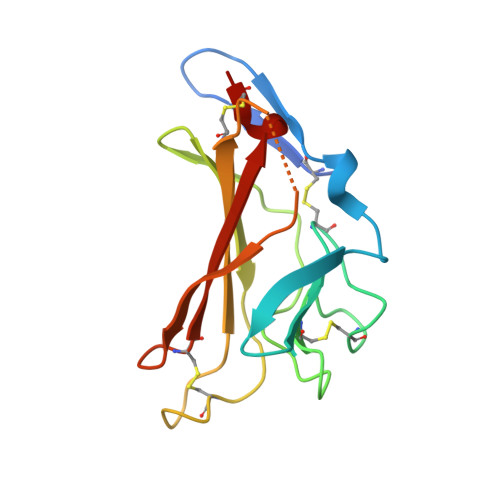
    Insulin-like growth factor II receptor (IGF2R) is a multifunctional cell surface receptor implicated in tumour suppression. Its growth inhibitory activity has been associated with an ability to bind IGF-II. IGF2R contains 15 homologous extracellular domains, with domain 11 primarily responsible for IGF-II binding. We report a 1.4 A resolution crystal structure of domain 11, solved using the anomalous scattering signal of sulfur. The structure consists of two crossed beta-sheets forming a flattened beta-barrel. Structural analysis identifies the putative IGF-II binding site at one end of the beta-barrel whilst crystal lattice contacts suggest a model for the full-length IGF2R extracellular region. The structure factors and coordinates of IGF2R domain 11 have been deposited in the Protein Data Bank (accession codes 1GP0 and 1GP3).


  • Organizational Affiliation
    • Cancer Research UK Receptor Structure Research Group, Division of Structural Biology, Wellcome Trust Centre for Human Genetics, University of Oxford, Roosevelt Drive, Headington, Oxford OX3 7BN, UK.

Macromolecule Content 

  • Total Structure Weight: 15.56 kDa 
  • Atom Count: 1,061 
  • Modeled Residue Count: 129 
  • Deposited Residue Count: 143 
  • Unique protein chains: 1

Macromolecules

Find similar proteins by:|  3D Structure
Entity ID: 1
MoleculeChains  Sequence LengthOrganismDetailsImage
CATION-INDEPENDENT MANNOSE-6-PHOSPHATE RECEPTOR143Homo sapiensMutation(s): 0 
UniProt & NIH Common Fund Data Resources
Find proteins for P11717 (Homo sapiens)
Explore P11717 
Go to UniProtKB:  P11717
PHAROS:  P11717
GTEx:  ENSG00000197081 
Entity Groups
Sequence Clusters30% Identity50% Identity70% Identity90% Identity95% Identity100% Identity
UniProt GroupP11717
Sequence Annotations
Expand
Reference Sequence

Experimental Data & Validation

Experimental Data

  • Method: X-RAY DIFFRACTION
  • Resolution: 1.95 Å
  • R-Value Free:  0.255 (Depositor), 0.258 (DCC) 
  • R-Value Work:  0.206 (Depositor), 0.206 (DCC) 
  • R-Value Observed: 0.206 (Depositor) 
Space Group: P 21 21 21
Unit Cell:
Length ( Å )Angle ( ˚ )
a = 31.373α = 90
b = 49.843β = 90
c = 83.03γ = 90
Software Package:
Software NamePurpose
CNSrefinement
DENZOdata reduction
SCALEPACKdata scaling
EPMRphasing

Structure Validation

View Full Validation Report


Entry History 

Deposition Data

Revision History  (Full details and data files)

  • Version 1.0: 2002-02-28
    Type: Initial release
  • Version 1.1: 2011-05-08
    Changes: Version format compliance
  • Version 1.2: 2011-07-13
    Changes: Version format compliance
  • Version 1.3: 2019-04-10
    Changes: Data collection, Other, Source and taxonomy
  • Version 1.4: 2023-12-13
    Changes: Data collection, Database references, Other, Refinement description
  • Version 1.5: 2024-11-06
    Changes: Structure summary