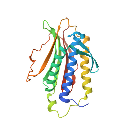
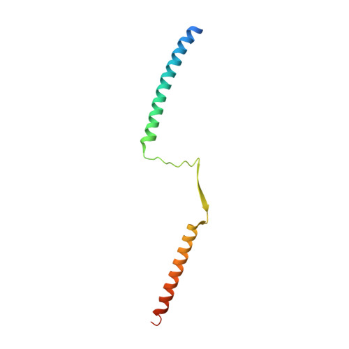
1GO4

Crystal structure of Mad1-Mad2 reveals a conserved Mad2 binding motif in Mad1 and Cdc20.


Experimental Data Snapshot

  • Method: X-RAY DIFFRACTION
  • Resolution: 2.05 Å
  • R-Value Free: 0.268 
  • R-Value Work: 0.240 
  • R-Value Observed: 0.240 

Starting Model: experimental
View more details

wwPDB Validation   3D Report Full Report


This is version 1.7 of the entry. See complete history


Literature

Crystal structure of the tetrameric Mad1-Mad2 core complex: implications of a 'safety belt' binding mechanism for the spindle checkpoint.

Sironi, L.Mapelli, M.Knapp, S.De Antoni, A.Jeang, K.T.Musacchio, A.

(2002) EMBO J 21: 2496-2506

  • DOI: https://doi.org/10.1093/emboj/21.10.2496
  • Primary Citation of Related Structures:  
    1GO4

  • PubMed Abstract: 

    The spindle checkpoint protein Mad1 recruits Mad2 to unattached kinetochores and is essential for Mad2-Cdc20 complex formation in vivo but not in vitro. The crystal structure of the Mad1-Mad2 complex reveals an asymmetric tetramer, with elongated Mad1 monomers parting from a coiled-coil to form two connected sub-complexes with Mad2. The Mad2 C-terminal tails are hinged mobile elements wrapping around the elongated ligands like molecular 'safety belts'. We show that Mad1 is a competitive inhibitor of the Mad2-Cdc20 complex, and propose that the Mad1-Mad2 complex acts as a regulated gate to control Mad2 release for Cdc20 binding. Mad1-Mad2 is strongly stabilized in the tetramer, but a 1:1 Mad1-Mad2 complex slowly releases Mad2 for Cdc20 binding, driven by favourable binding energies. Thus, the rate of Mad2 binding to Cdc20 during checkpoint activation may be regulated by conformational changes that destabilize the tetrameric Mad1-Mad2 assembly to promote Mad2 release. We also show that unlocking the Mad2 C-terminal tail is required for ligand release from Mad2, and that the 'safety belt' mechanism may prolong the lifetime of Mad2-ligand complexes.


  • Organizational Affiliation

    Structural Biology Unit, Department of Experimental Oncology, European Institute of Oncology, Via Ripamonti 435, 20141 Milan, Italy.


Macromolecules
Find similar proteins by:  (by identity cutoff)  |  3D Structure
Entity ID: 1
MoleculeChains Sequence LengthOrganismDetailsImage
Mitotic spindle assembly checkpoint protein MAD2A
A, B, C, D
205Homo sapiensMutation(s): 1 
Gene Names: MAD2L1MAD2
UniProt & NIH Common Fund Data Resources
Find proteins for Q13257 (Homo sapiens)
Explore Q13257 
Go to UniProtKB:  Q13257
PHAROS:  Q13257
GTEx:  ENSG00000164109 
Entity Groups  
Sequence Clusters30% Identity50% Identity70% Identity90% Identity95% Identity100% Identity
UniProt GroupQ13257
Sequence Annotations
Expand
  • Reference Sequence
Find similar proteins by:  (by identity cutoff)  |  3D Structure
Entity ID: 2
MoleculeChains Sequence LengthOrganismDetailsImage
Mitotic spindle assembly checkpoint protein MAD1
E, F, G, H
100Homo sapiensMutation(s): 0 
Gene Names: MAD1L1MAD1TXBP181
UniProt & NIH Common Fund Data Resources
Find proteins for Q9Y6D9 (Homo sapiens)
Explore Q9Y6D9 
Go to UniProtKB:  Q9Y6D9
PHAROS:  Q9Y6D9
GTEx:  ENSG00000002822 
Entity Groups  
Sequence Clusters30% Identity50% Identity70% Identity90% Identity95% Identity100% Identity
UniProt GroupQ9Y6D9
Sequence Annotations
Expand
  • Reference Sequence
Experimental Data & Validation

Experimental Data

  • Method: X-RAY DIFFRACTION
  • Resolution: 2.05 Å
  • R-Value Free: 0.268 
  • R-Value Work: 0.240 
  • R-Value Observed: 0.240 
  • Space Group: P 1 21 1
Unit Cell:
Length ( Å )Angle ( ˚ )
a = 111.037α = 90
b = 63.023β = 111.65
c = 139.511γ = 90
Software Package:
Software NamePurpose
CNSrefinement
DENZOdata reduction
SCALEPACKdata scaling
SnBphasing

Structure Validation

View Full Validation Report


Entry History 

Deposition Data

Revision History  (Full details and data files)

  • Version 1.0: 2002-05-16
    Type: Initial release
  • Version 1.1: 2011-05-08
    Changes: Version format compliance
  • Version 1.2: 2011-07-13
    Changes: Version format compliance
  • Version 1.3: 2017-07-05
    Changes: Refinement description
  • Version 1.4: 2018-06-20
    Changes: Data collection, Database references, Source and taxonomy, Structure summary
  • Version 1.5: 2019-05-08
    Changes: Data collection, Experimental preparation
  • Version 1.6: 2019-10-16
    Changes: Data collection
  • Version 1.7: 2023-12-13
    Changes: Data collection, Database references, Refinement description