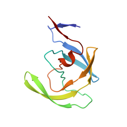
Crystal structures of complexes of a peptidic inhibitor with wild-type and two mutant HIV-1 proteases.
Hong, L., Treharne, A., Hartsuck, J.A., Foundling, S., Tang, J.(1996) Biochemistry 35: 10627-10633
- PubMed: 8718851
- DOI: https://doi.org/10.1021/bi960481s
- Primary Citation of Related Structures:
1GNM, 1GNN, 1GNO - PubMed Abstract:
Crystal structures of the protease of human immunodeficiency virus type 1 (HIV-1) and two mutant proteases, V82D and V82N, have been determined. In all three cases the enzyme forms a complex with the peptidic inhibitor U-89360E. All structures have been determined to 2.3 A resolution and have satisfactory agreement factors: 0.173 for wild type, 0.175 for V82D, and 0.182 for V82N. Comparison of the three crystal structures provides explanations which are consistent with the known kinetic properties of these mutant enzymes with the U-89360E inhibitor [Lin, Y., Lin, X., Hong, L., Foundling, S., Heinrikson, R. L., Thaisrivongs, S., Leelamanit, W., Raterman, D., Shah, M., Dunn, B.M., & Tang, J. (1995) Biochemistry 34, 1143-1152]. Unfavorable van der Waals interactions between the inhibitor and the mutated side chains at position 82 are consistent with diminished affinity for the inhibitor by the mutant enzymes. If a mutation is potentially resistant to an inhibitor, the mutant enzyme should not only have an increased Ki for the inhibitor but should also preserve considerable catalytic capability. The V82D mutant possesses these qualities. In the V82D crystal structure, a water molecule, which connects the protease flap to the inhibitor, is missing or of low occupancy. Absence of this bridge may be important in determining catalytic capability. Moreover, mutation at position 82 induces change in two polypeptide backbone regions, 35-41 and 67-68, which may be related to protease flap mobility.
Organizational Affiliation:
Protein Studies Program, Oklahoma Medical Research Foundation, University of Oklahoma Health Sciences Center, Oklahoma City 73104, USA.


