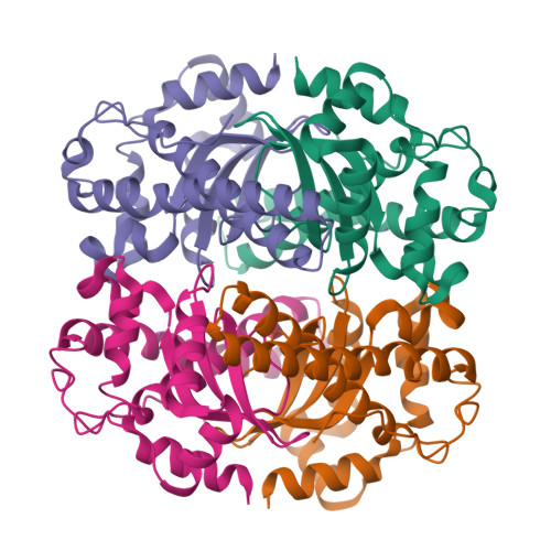
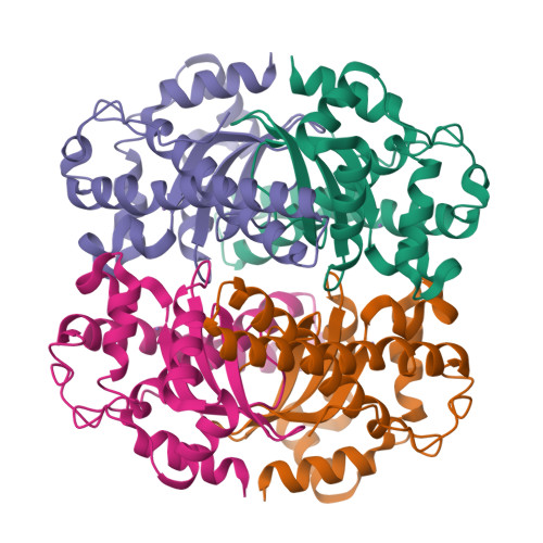
Engineering a Change in the Metal-Ion Specificity of the Iron-Depedent Superoxide Dismutase from Mycobacterium Tuberculosis. X-Ray Structure Analysis of Site-Directed Mutants
Bunting, K.A., Cooper, J.B., Badasso, M.O., Tickle, I.J., Newton, M., Wood, S.P., Young, D.B.(1998) Eur J Biochem 251: 795
- PubMed: 9490054
- DOI: https://doi.org/10.1046/j.1432-1327.1998.2510795.x
- Primary Citation of Related Structures:
1GN3, 1GN4 - PubMed Abstract:
We have refined the X-ray structures of two site-directed mutants of the iron-dependent superoxide dismutase (SOD) from Mycobacterium tuberculosis. These mutations which affect residue 145 in the enzyme (H145Q and H145E) were designed to alter its metal-ion specificity. This residue is either Gln or His in homologous SOD enzymes and has previously been shown to play a role in active-site interactions since its side-chain helps to coordinate the metal ion via a solvent molecule which is thought to be a hydroxide ion. The mutations were based on the observation that in the closely homologous manganese dependent SOD from Mycobacterium leprae, the only significant difference from the M. tuberculosis SOD within 10 A of the metal-binding site is the substitution of Gln for His at position 145. Hence an H145Q mutant of the M. tuberculosis (TB) SOD was engineered to investigate this residue's role in metal ion dependence and an isosteric H145E mutant was also expressed. The X-ray structures of the H145Q and H145E mutants have been solved at resolutions of 4.0 A and 2.5 A, respectively, confirming that neither mutation has any gross effects on the conformation of the enzyme or the structure of the active site. The residue substitutions are accommodated in the enzyme's three-dimensional structure by small local conformational changes. Peroxide inhibition experiments and atomic absorption spectroscopy establish surprisingly the H145E mutant SOD has manganese bound to it whereas the H145Q mutant SOD retains iron as the active-site metal. This alteration in metal specificity may reflect on the preference of manganese ions for anionic ligands.
Organizational Affiliation:
Department of Crystallography, Birkbeck College, University of London, England.


