1GK4 | pdb_00001gk4

HUMAN VIMENTIN COIL 2B FRAGMENT (CYS2)


Experimental Data Snapshot

  • Method: X-RAY DIFFRACTION
  • Resolution: 2.30 Å
  • R-Value Free: 
    0.262 (Depositor), 0.263 (DCC) 
  • R-Value Work: 
    0.242 (Depositor), 0.241 (DCC) 
  • R-Value Observed: 
    0.242 (Depositor) 

wwPDB Validation 3D Report Full Report

Validation slider image for 1GK4

This is version 1.3 of the entry. See complete history

Literature

Conserved Segments 1A and 2B of the Intermediate Filament Dimer: Their Atomic Structures and Role in Filament Assembly.

Strelkov, S.Herrmann, H.Geisler, N.Wedig, T.Zimbelmann, R.Aebi, U.Burkhard, P.

(2002) EMBO J 21: 1255

  • DOI: https://doi.org/10.1093/emboj/21.6.1255
  • Primary Citation Related Structures: 
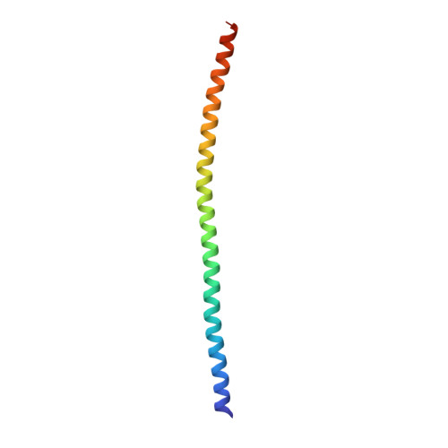
    1GK4, 1GK6, 1GK7

  • PubMed Abstract: 

    Intermediate filaments (IFs) are key components of the cytoskeleton in higher eukaryotic cells. The elementary IF 'building block' is an elongated coiled-coil dimer consisting of four consecutive alpha-helical segments. The segments 1A and 2B include highly conserved sequences and are critically involved in IF assembly. Based on the crystal structures of three human vimentin fragments at 1.4-2.3 A resolution (PDB entries 1gk4, 1gk6 and 1gk7), we have established the molecular organization of these two segments. The fragment corresponding to segment 1A forms a single, amphipatic alpha-helix, which is compatible with a coiled-coil geometry. While this segment might yield a coiled coil within an isolated dimer, monomeric 1A helices are likely to play a role in specific dimer-dimer interactions during IF assembly. The 2B segment reveals a double-stranded coiled coil, which unwinds near residue Phe351 to accommodate a 'stutter'. A fragment containing the last seven heptads of 2B interferes heavily with IF assembly and also transforms mature vimentin filaments into a new kind of structure. These results provide the first insight into the architecture and functioning of IFs at the atomic level.


  • Organizational Affiliation
    • Maurice E.Müller Institute for Structural Biology, Biozentrum, University of Basel, Klingelbergstrasse 70, CH-4056 Basel, Switzerland. sergei-v.strelkov@unibas.ch

Macromolecule Content 

  • Total Structure Weight: 59.58 kDa 
  • Atom Count: 4,169 
  • Modeled Residue Count: 450 
  • Deposited Residue Count: 504 
  • Unique protein chains: 1

Macromolecules

Find similar proteins by:|  3D Structure
Entity ID: 1
MoleculeChains  Sequence LengthOrganismDetailsImage
VIMENTIN
A, B, C, D, E
A, B, C, D, E, F
84Homo sapiensMutation(s): 0 
UniProt & NIH Common Fund Data Resources
Find proteins for P08670 (Homo sapiens)
Explore P08670 
Go to UniProtKB:  P08670
PHAROS:  P08670
GTEx:  ENSG00000026025 
Entity Groups
Sequence Clusters30% Identity50% Identity70% Identity90% Identity95% Identity100% Identity
UniProt GroupP08670
Sequence Annotations
Expand
Reference Sequence

Small Molecules

Ligands 1 Unique
IDChains Name / Formula / InChI Key2D Diagram3D Interactions
ACT

Query on ACT


Download:Ideal Coordinates CCD File
G [auth B]ACETATE ION
C2 H3 O2
QTBSBXVTEAMEQO-UHFFFAOYSA-M

Experimental Data & Validation

Experimental Data

  • Method: X-RAY DIFFRACTION
  • Resolution: 2.30 Å
  • R-Value Free:  0.262 (Depositor), 0.263 (DCC) 
  • R-Value Work:  0.242 (Depositor), 0.241 (DCC) 
  • R-Value Observed: 0.242 (Depositor) 
Space Group: I 2 2 2
Unit Cell:
Length ( Å )Angle ( ˚ )
a = 76.594α = 90
b = 84.283β = 90
c = 240.785γ = 90
Software Package:
Software NamePurpose
CNSrefinement
DENZOdata reduction
SCALEPACKdata scaling
SHARPphasing

Structure Validation

View Full Validation Report


Entry History 

Deposition Data

Revision History  (Full details and data files)

  • Version 1.0: 2002-03-15
    Type: Initial release
  • Version 1.1: 2011-05-08
    Changes: Version format compliance
  • Version 1.2: 2011-07-13
    Changes: Version format compliance
  • Version 1.3: 2024-05-08
    Changes: Data collection, Database references, Other