1GJR | pdb_00001gjr

Ferredoxin-NADP+ Reductase complexed with NADP+ by COCRYSTALLIZATION


Experimental Data Snapshot

  • Method: X-RAY DIFFRACTION
  • Resolution: 2.10 Å
  • R-Value Free: 
    0.270 (Depositor), 0.259 (DCC) 
  • R-Value Work: 
    0.200 (Depositor), 0.197 (DCC) 
  • R-Value Observed: 
    0.200 (Depositor) 

Starting Model: experimental
View more details

wwPDB Validation 3D Report Full Report

Validation slider image for 1GJR

Ligand Structure Quality Assessment 


This is version 1.4 of the entry. See complete history

Literature

Mechanism of Coenzyme Recognition and Binding Revealed by Crystal Structure Analysis of Ferredoxin-Nadp(+) Reductase Complexed with Nadp(+)

Hermoso, J.Mayoral, T.Faro, M.Gomez-Moreno, C.Sanz-Aparicio, J.Medina, M.

(2002) J Mol Biology 319: 1133

  • DOI: https://doi.org/10.1016/S0022-2836(02)00388-1
  • Primary Citation Related Structures: 
    1GJR

  • PubMed Abstract: 

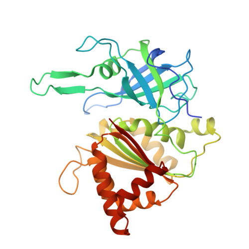
    The flavoenzyme ferredoxin-NADP+ reductase (FNR) catalyses the production of NADPH in photosynthesis. The three-dimensional structure of FNR presents two distinct domains, one for binding of the FAD prosthetic group and the other for NADP+ binding. In spite of extensive experiments and different crystallographic approaches, many aspects about how the NADP+ substrate binds to FNR and how the hydride ion is transferred from FAD to NADP+ remain unclear. The structure of an FNR:NADP+ complex from Anabaena has been determined by X-ray diffraction analysis of the cocrystallised units to 2.1 A resolution. Structural perturbation of FNR induced by complex formation produces a narrower cavity in which the 2'-phospho-AMP and pyrophosphate portions of the NADP+ are perfectly bound. In addition, the nicotinamide mononucleotide moiety is placed in a new pocket created near the FAD cofactor with the ribose being in a tight conformation. The crystal structure of this FNR:NADP+ complex obtained by cocrystallisation displays NADP+ in an unusual conformation and can be considered as an intermediate state in the process of coenzyme recognition and binding. Structural analysis and comparison with previously reported complexes allow us to postulate a mechanism which would permit efficient hydride transfer to occur. Besides, this structure gives new insights into the postulated formation of the ferredoxin:FNR:NADP+ ternary complex by prediction of new intermolecular interactions, which could only exist after FNR:NADP+ complex formation. Finally, structural comparison with the members of the broad FNR structural family also provides an explanation for the high specificity exhibited by FNR for NADP+/H versus NAD+/H.


  • Organizational Affiliation
    • Grupo de Cristalografía Macromolecular y Biología Estructural, Instituto Química-Física Rocasolano, C.S.I.C., Serrano 119, 28006 Madrid, Spain. xjuan@iqfr.csic.es

Macromolecule Content 

  • Total Structure Weight: 35.7 kDa 
  • Atom Count: 2,639 
  • Modeled Residue Count: 295 
  • Deposited Residue Count: 304 
  • Unique protein chains: 1

Macromolecules

Find similar proteins by:|  3D Structure
Entity ID: 1
MoleculeChains  Sequence LengthOrganismDetailsImage
FERREDOXIN-NADP REDUCTASE304Nostoc sp. PCC 7119Mutation(s): 0 
EC: 1.18.1.2
UniProt
Find proteins for P21890 (Nostoc sp. (strain ATCC 29151 / PCC 7119))
Explore P21890 
Go to UniProtKB:  P21890
Entity Groups
Sequence Clusters30% Identity50% Identity70% Identity90% Identity95% Identity100% Identity
UniProt GroupP21890
Sequence Annotations
Expand
Reference Sequence

Small Molecules

Ligands 2 Unique
IDChains Name / Formula / InChI Key2D Diagram3D Interactions
FAD

Query on FAD


Download:Ideal Coordinates CCD File
B [auth A]FLAVIN-ADENINE DINUCLEOTIDE
C27 H33 N9 O15 P2
VWWQXMAJTJZDQX-UYBVJOGSSA-N
NAP

Query on NAP


Download:Ideal Coordinates CCD File
C [auth A]NADP NICOTINAMIDE-ADENINE-DINUCLEOTIDE PHOSPHATE
C21 H28 N7 O17 P3
XJLXINKUBYWONI-NNYOXOHSSA-N

Experimental Data & Validation

Experimental Data

  • Method: X-RAY DIFFRACTION
  • Resolution: 2.10 Å
  • R-Value Free:  0.270 (Depositor), 0.259 (DCC) 
  • R-Value Work:  0.200 (Depositor), 0.197 (DCC) 
  • R-Value Observed: 0.200 (Depositor) 
Space Group: I 41
Unit Cell:
Length ( Å )Angle ( ˚ )
a = 151.69α = 90
b = 151.69β = 90
c = 35.44γ = 90
Software Package:
Software NamePurpose
X-PLORrefinement
MOSFLMdata reduction
SCALAdata scaling
AMoREphasing

Structure Validation

View Full Validation Report


Ligand Structure Quality Assessment 


Entry History 

Deposition Data

Revision History  (Full details and data files)

  • Version 1.0: 2002-06-27
    Type: Initial release
  • Version 1.1: 2011-05-08
    Changes: Version format compliance
  • Version 1.2: 2011-07-13
    Changes: Version format compliance
  • Version 1.3: 2019-05-22
    Changes: Data collection, Other, Refinement description
  • Version 1.4: 2023-12-13
    Changes: Data collection, Database references, Derived calculations, Refinement description