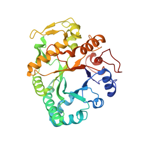
Three-dimensional structures of two plant beta-glucan endohydrolases with distinct substrate specificities.
Varghese, J.N., Garrett, T.P., Colman, P.M., Chen, L., Hoj, P.B., Fincher, G.B.(1994) Proc Natl Acad Sci U S A 91: 2785-2789
- PubMed: 8146192
- DOI: https://doi.org/10.1073/pnas.91.7.2785
- Primary Citation of Related Structures:
1GHR, 1GHS - PubMed Abstract:
The three-dimensional structures of (1-->3)-beta-glucanase (EC 3.2.1.39) isoenzyme GII and (1-->3,1-->4)-beta-glucanase (EC 3.2.1.73) isoenzyme EII from barley have been determined by x-ray crystallography at 2.2- to 2.3-A resolution. The two classes of polysaccharide endohydrolase differ in their substrate specificity and function. Thus, the (1-->3)-beta-glucanases, which are classified amongst the plant "pathogenesis-related proteins," can hydrolyze (1-->3)- and (1-->3,1-->6)-beta-glucans of fungal cell walls and may therefore contribute to plant defense strategies, while the (1-->3,1-->4)-beta-glucanases function in plant cell wall hydrolysis during mobilization of the endosperm in germinating grain or during the growth of vegetative tissues. Both enzymes are alpha/beta-barrel structures. The catalytic amino acid residues are located within deep grooves which extend across the enzymes and which probably bind the substrates. Because the polypeptide backbones of the two enzymes are structurally very similar, the differences in their substrate specificities, and hence their widely divergent functions, have been acquired primarily by amino acid substitutions within the groove.
Organizational Affiliation:
Biomolecular Research Institute, Parkville, Victoria, Australia.


