1GEC | pdb_00001gec

GLYCYL ENDOPEPTIDASE-COMPLEX WITH BENZYLOXYCARBONYL-LEUCINE-VALINE-GLYCINE-METHYLENE COVALENTLY BOUND TO CYSTEINE 25


Experimental Data Snapshot

  • Method: X-RAY DIFFRACTION
  • Resolution: 2.10 Å
  • R-Value Free: 
    0.258 (Depositor) 
  • R-Value Work: 
    0.196 (Depositor) 
  • R-Value Observed: 
    0.196 (Depositor) 

wwPDB Validation 3D Report Full Report

Validation slider image for 1GEC

This is version 1.4 of the entry. See complete history

Literature

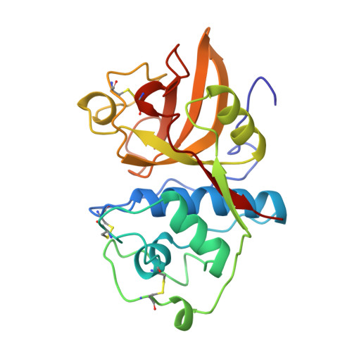
Crystal structure of glycyl endopeptidase from Carica papaya: a cysteine endopeptidase of unusual substrate specificity.

O'Hara, B.P.Hemmings, A.M.Buttle, D.J.Pearl, L.H.

(1995) Biochemistry 34: 13190-13195

  • DOI: https://doi.org/10.1021/bi00040a034
  • Primary Citation Related Structures: 
    1GEC

  • PubMed Abstract: 

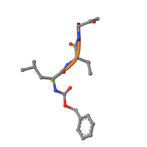
    Glycyl endopeptidase is a cysteine endopeptidase of the papain family, characterized by specificity for cleavage C-terminal to glycyl residues only and by resistance to inhibition by members of the cystatin family of cysteine proteinase inhibitors. Glycyl endopeptidase has been crystallized from high salt with a substrate-like inhibitor covalently bound to the catalytic Cys 25. The structure has been solved by molecular replacement with the structure of papain and refined at 2.1 A to an R factor of 0.196 (Rfree = 0.258) with good geometry. The structure of the S1 substrate binding site of glycyl endopeptidase differs from that of papain by the substitution of glycines at residues 23 and 65 in papain, with glutamic acid and arginine, respectively, in glycyl endopeptidase. The side chains of these residues form a barrier across the binding pocket, effectively excluding substrate residues with large side chains from the S1 subsite. The constriction of this subsite in glycyl endopeptidase explains the unique specificity of this enzyme for cleavage after glycyl residues and is a major component of its resistance to inhibition by cystatins.


  • Organizational Affiliation
    • Department of Biochemistry and Molecular Biology, University College London, U.K.

Macromolecule Content 

  • Total Structure Weight: 23.81 kDa 
  • Atom Count: 1,789 
  • Modeled Residue Count: 221 
  • Deposited Residue Count: 221 
  • Unique protein chains: 2

Macromolecules

Find similar proteins by:|  3D Structure
Entity ID: 1
MoleculeChains  Sequence LengthOrganismDetailsImage
GLYCYL ENDOPEPTIDASEA [auth E]216Carica papayaMutation(s): 0 
EC: 3.4.22.25
UniProt
Find proteins for P05994 (Carica papaya)
Explore P05994 
Go to UniProtKB:  P05994
Entity Groups
Sequence Clusters30% Identity50% Identity70% Identity90% Identity95% Identity100% Identity
UniProt GroupP05994
Sequence Annotations
Expand
Reference Sequence
Find similar proteins by:  Sequence   |   3D Structure  
Entity ID: 2
MoleculeChains  Sequence LengthOrganismDetailsImage
BENZYLOXYCARBONYL-LEUCINE-VALINE-GLYCINE-METHYLENE INHIBITORB [auth I]5N/AMutation(s): 0 

Biologically Interesting Molecules (External Reference) 

1 Unique

Experimental Data & Validation

Experimental Data

  • Method: X-RAY DIFFRACTION
  • Resolution: 2.10 Å
  • R-Value Free:  0.258 (Depositor) 
  • R-Value Work:  0.196 (Depositor) 
  • R-Value Observed: 0.196 (Depositor) 
Space Group: P 31
Unit Cell:
Length ( Å )Angle ( ˚ )
a = 55.78α = 90
b = 55.78β = 90
c = 64.43γ = 120
Software Package:
Software NamePurpose
MOSFLMdata reduction
X-PLORmodel building
X-PLORrefinement
X-PLORphasing

Structure Validation

View Full Validation Report


Entry History 

Deposition Data

Revision History  (Full details and data files)

  • Version 1.0: 1995-12-07
    Type: Initial release
  • Version 1.1: 2008-03-24
    Changes: Version format compliance
  • Version 1.2: 2011-07-13
    Changes: Atomic model, Database references, Derived calculations, Non-polymer description, Structure summary, Version format compliance
  • Version 1.3: 2012-12-12
    Changes: Other
  • Version 1.4: 2024-10-30
    Changes: Data collection, Database references, Derived calculations, Structure summary