1G83 | pdb_00001g83

CRYSTAL STRUCTURE OF FYN SH3-SH2


Experimental Data Snapshot

  • Method: X-RAY DIFFRACTION
  • Resolution: 2.60 Å
  • R-Value Free: 
    0.277 (Depositor), 0.292 (DCC) 
  • R-Value Work: 
    0.214 (Depositor), 0.228 (DCC) 

wwPDB Validation 3D Report Full Report

Validation slider image for 1G83

This is version 1.4 of the entry. See complete history

Literature

The role of the Src homology 3-Src homology 2 interface in the regulation of Src kinases.

Arold, S.T.Ulmer, T.S.Mulhern, T.D.Werner, J.M.Ladbury, J.E.Campbell, I.D.Noble, M.E.

(2001) J Biological Chem 276: 17199-17205

  • DOI: https://doi.org/10.1074/jbc.M011185200
  • Primary Citation Related Structures: 
    1G83

  • PubMed Abstract: 

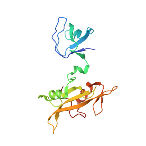
    The regulatory fragment of Src kinases, comprising Src homology (SH) 3 and SH2 domains, is responsible for controlled repression of kinase activity. We have used a multidisciplinary approach involving crystallography, NMR, and isothermal titration calorimetry to study the regulatory fragment of Fyn (FynSH32) and its interaction with a physiological activator: a fragment of focal adhesion kinase that contains both phosphotyrosine and polyproline motifs. Although flexible, the preferred disposition of SH3 and SH2 domains in FynSH32 resembles the inactive forms of Hck and Src, differing significantly from LckSH32. This difference, which results from variation in the SH3-SH2 linker sequences, will affect the potential of the regulatory fragments to repress kinase activity. This surprising result implies that the mechanism of repression of Src family members may vary, explaining functional distinctions between Fyn and Lck. The interaction between FynSH32 and focal adhesion kinase is restricted to the canonical SH3 and SH2 binding sites and does not affect the dynamic independence of the two domains. Consequently, the interaction shows no enhancement by an avidity effect. Such an interaction may have evolved to gain specificity through an extended recognition site while maintaining rapid dissociation after signaling.


  • Organizational Affiliation
    • Laboratory of Molecular Biophysics and Department of Biochemistry, University of Oxford, South Parks Road, Oxford OX1 3QU, United Kingdom.

Macromolecule Content 

  • Total Structure Weight: 37.82 kDa 
  • Atom Count: 2,642 
  • Modeled Residue Count: 322 
  • Deposited Residue Count: 330 
  • Unique protein chains: 1

Macromolecules

Find similar proteins by:|  3D Structure
Entity ID: 1
MoleculeChains  Sequence LengthOrganismDetailsImage
PROTO-ONCOGENE TYROSINE-PROTEIN KINASE FYN
A, B
165Homo sapiensMutation(s): 3 
Gene Names: FYN
EC: 2.7.1.112 (PDB Primary Data), 2.7.10.2 (UniProt)
UniProt & NIH Common Fund Data Resources
Find proteins for P06241 (Homo sapiens)
Explore P06241 
Go to UniProtKB:  P06241
PHAROS:  P06241
GTEx:  ENSG00000010810 
Entity Groups
Sequence Clusters30% Identity50% Identity70% Identity90% Identity95% Identity100% Identity
UniProt GroupP06241
Sequence Annotations
Expand
Reference Sequence

Experimental Data & Validation

Experimental Data

  • Method: X-RAY DIFFRACTION
  • Resolution: 2.60 Å
  • R-Value Free:  0.277 (Depositor), 0.292 (DCC) 
  • R-Value Work:  0.214 (Depositor), 0.228 (DCC) 
Space Group: P 1 21 1
Unit Cell:
Length ( Å )Angle ( ˚ )
a = 39.97α = 90
b = 89.96β = 101.44
c = 60.27γ = 90
Software Package:
Software NamePurpose
AMoREphasing
CNSrefinement
MOSFLMdata reduction
CCP4data scaling

Structure Validation

View Full Validation Report


Entry History 

Deposition Data

Revision History  (Full details and data files)

  • Version 1.0: 2001-05-30
    Type: Initial release
  • Version 1.1: 2008-04-27
    Changes: Version format compliance
  • Version 1.2: 2011-07-13
    Changes: Version format compliance
  • Version 1.3: 2021-10-27
    Changes: Database references
  • Version 1.4: 2024-02-07
    Changes: Data collection