1G6N | pdb_00001g6n

2.1 ANGSTROM STRUCTURE OF CAP-CAMP


Experimental Data Snapshot

  • Method: X-RAY DIFFRACTION
  • Resolution: 2.10 Å
  • R-Value Work: 
    0.207 (Depositor) 
  • R-Value Observed: 
    0.207 (Depositor) 

Starting Model: experimental
View more details

wwPDB Validation 3D Report Full Report

Validation slider image for 1G6N

This is version 1.4 of the entry. See complete history

Literature

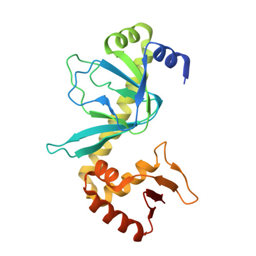
Modeling the cAMP-induced allosteric transition using the crystal structure of CAP-cAMP at 2.1 A resolution.

Passner, J.M.Schultz, S.C.Steitz, T.A.

(2000) J Mol Biology 304: 847-859

  • DOI: https://doi.org/10.1006/jmbi.2000.4231
  • Primary Citation Related Structures: 
    1G6N

  • PubMed Abstract: 

    After an allosteric transition produced by the binding of cyclic AMP (cAMP), the Escherichia coli catabolite gene activator protein (CAP) binds DNA specifically and activates transcription. The three-dimensional crystal structure of the CAP-cAMP complex has been refined at 2.1 A resolution, thus enabling a better evaluation of the structural basis for CAP phenotypes, the interactions of cAMP with CAP and the roles played by water structure. A review of mutational analysis of CAP together with the additional structural information presented here suggests a possible mechanism for the cAMP-induced allostery required for DNA binding and transcriptional activation. We hypothesize that cAMP binding may reorient the coiled-coil C-helices, which provide most of the dimer interface, thereby altering the relative positions of the DNA-binding domains of the CAP dimer. Additionally, cAMP binding may cause a further rearrangement of the DNA-binding and cAMP-binding domains of CAP via a flap consisting of beta-strands 4 and 5 which lies over the cAMP.


  • Organizational Affiliation
    • Department of Molecular Biophysics and Biochemistry, Mount Sinai Schoolof Medicine, New York, NY 10029, USA. Passner@inka.mssm.edu

Macromolecule Content 

  • Total Structure Weight: 48 kDa 
  • Atom Count: 3,329 
  • Modeled Residue Count: 401 
  • Deposited Residue Count: 420 
  • Unique protein chains: 1

Macromolecules

Find similar proteins by:|  3D Structure
Entity ID: 1
MoleculeChains  Sequence LengthOrganismDetailsImage
CATABOLITE GENE ACTIVATOR PROTEIN
A, B
210Escherichia coliMutation(s): 0 
UniProt
Find proteins for P0ACJ8 (Escherichia coli (strain K12))
Explore P0ACJ8 
Go to UniProtKB:  P0ACJ8
Entity Groups
Sequence Clusters30% Identity50% Identity70% Identity90% Identity95% Identity100% Identity
UniProt GroupP0ACJ8
Sequence Annotations
Expand
Reference Sequence

Small Molecules

Ligands 1 Unique
IDChains Name / Formula / InChI Key2D Diagram3D Interactions
CMP

Query on CMP


Download:Ideal Coordinates CCD File
C [auth A],
D [auth B]
ADENOSINE-3',5'-CYCLIC-MONOPHOSPHATE
C10 H12 N5 O6 P
IVOMOUWHDPKRLL-KQYNXXCUSA-N

Experimental Data & Validation

Experimental Data

  • Method: X-RAY DIFFRACTION
  • Resolution: 2.10 Å
  • R-Value Work:  0.207 (Depositor) 
  • R-Value Observed: 0.207 (Depositor) 
Space Group: P 21 21 21
Unit Cell:
Length ( Å )Angle ( ˚ )
a = 46.8α = 90
b = 95.6β = 90
c = 105.5γ = 90
Software Package:
Software NamePurpose
X-PLORmodel building
X-PLORrefinement
UCSD-systemdata reduction
SCALEPACKdata scaling
X-PLORphasing

Structure Validation

View Full Validation Report


Entry History 

Deposition Data

Revision History  (Full details and data files)

  • Version 1.0: 2000-12-15
    Type: Initial release
  • Version 1.1: 2008-04-27
    Changes: Version format compliance
  • Version 1.2: 2011-07-13
    Changes: Version format compliance
  • Version 1.3: 2017-10-04
    Changes: Refinement description
  • Version 1.4: 2023-08-09
    Changes: Data collection, Database references, Derived calculations, Refinement description