1G3M | pdb_00001g3m

CRYSTAL STRUCTURE OF HUMAN ESTROGEN SULFOTRANSFERASE IN COMPLEX WITH IN-ACTIVE COFACTOR PAP AND 3,5,3',5'-TETRACHLORO-BIPHENYL-4,4'-DIOL


Experimental Data Snapshot

  • Method: X-RAY DIFFRACTION
  • Resolution: 1.70 Å
  • R-Value Free: 
    0.219 (Depositor) 
  • R-Value Work: 
    0.193 (Depositor) 
  • R-Value Observed: 
    0.193 (Depositor) 

Starting Model: experimental
View more details

wwPDB Validation 3D Report Full Report

Validation slider image for 1G3M

This is version 1.4 of the entry. See complete history

Literature

Crystallographic analysis of a hydroxylated polychlorinated biphenyl (OH-PCB) bound to the catalytic estrogen binding site of human estrogen sulfotransferase.

Shevtsov, S.Petrotchenko, E.V.Pedersen, L.C.Negishi, M.

(2003) Environ Health Perspect 111: 884-888

  • DOI: https://doi.org/10.1289/ehp.6056
  • Primary Citation Related Structures: 
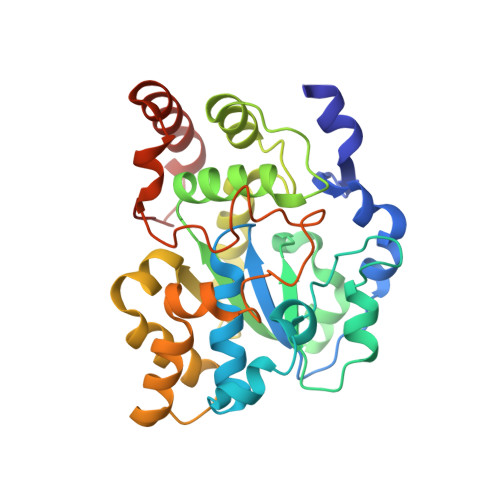
    1G3M

  • PubMed Abstract: 

    Certain hydroxylated polychlorinated biphenyls (OH-PCBs) inhibit the human estrogen sulfotransferase (hEST) at subnanomolar concentrations, suggesting a possible pathway for PCB toxicity due to environmental exposure in humans. To address the structural basis of the inhibition, we have determined the crystal structure of hEST in the presence of the sulfuryl donor product 3 -phosphoadenosine 5 -phosphate and the OH-PCB 4,4 -OH 3,5,3,5 -tetraCB. The OH-PCB binds in the estrogen binding site with the position of the first phenolic ring in an orientation similar to the phenolic ring of 17 beta-estradiol. Interestingly, the OH-PCB does not bind in a planar conformation, but rather with a 30-degree twist between the phenyl rings. The crystal structure of hEST with the OH-PCB bound gives physical evidence that certain OH-PCBs can mimic binding of estrogenic compounds in biological systems.


  • Organizational Affiliation
    • Pharmacogenetic Section, Laboratory of Reproductive and Developmental Toxicology, National Institute of Environmental Health Sciences, National Institutes of Health, Research Triangle Park, North Carolina 27709, USA.

Macromolecule Content 

  • Total Structure Weight: 72.1 kDa 
  • Atom Count: 5,484 
  • Modeled Residue Count: 575 
  • Deposited Residue Count: 588 
  • Unique protein chains: 1

Macromolecules

Find similar proteins by:|  3D Structure
Entity ID: 1
MoleculeChains  Sequence LengthOrganismDetailsImage
ESTROGEN SULFOTRANSFERASE
A, B
294Homo sapiensMutation(s): 0 
Gene Names: STE
EC: 2.8.2.4
UniProt & NIH Common Fund Data Resources
Find proteins for P49888 (Homo sapiens)
Explore P49888 
Go to UniProtKB:  P49888
PHAROS:  P49888
GTEx:  ENSG00000109193 
Entity Groups
Sequence Clusters30% Identity50% Identity70% Identity90% Identity95% Identity100% Identity
UniProt GroupP49888
Sequence Annotations
Expand
Reference Sequence

Small Molecules

Ligands 3 Unique
IDChains Name / Formula / InChI Key2D Diagram3D Interactions
A3P

Query on A3P


Download:Ideal Coordinates CCD File
C [auth A],
I [auth B]
ADENOSINE-3'-5'-DIPHOSPHATE
C10 H15 N5 O10 P2
WHTCPDAXWFLDIH-KQYNXXCUSA-N
PCQ

Query on PCQ


Download:Ideal Coordinates CCD File
D [auth A],
J [auth B]
3,5,3',5'-TETRACHLORO-BIPHENYL-4,4'-DIOL
C12 H6 Cl4 O2
YCYDXOVJXVALHY-UHFFFAOYSA-N
EDO

Query on EDO


Download:Ideal Coordinates CCD File
E [auth A],
F [auth A],
G [auth A],
H [auth A]
1,2-ETHANEDIOL
C2 H6 O2
LYCAIKOWRPUZTN-UHFFFAOYSA-N

Experimental Data & Validation

Experimental Data

  • Method: X-RAY DIFFRACTION
  • Resolution: 1.70 Å
  • R-Value Free:  0.219 (Depositor) 
  • R-Value Work:  0.193 (Depositor) 
  • R-Value Observed: 0.193 (Depositor) 
Space Group: P 1 21 1
Unit Cell:
Length ( Å )Angle ( ˚ )
a = 62.687α = 90
b = 96.894β = 92.58
c = 61.724γ = 90
Software Package:
Software NamePurpose
DENZOdata reduction
SCALEPACKdata scaling
CCP4model building
CNSrefinement
Omodel building
CCP4phasing
CNSphasing

Structure Validation

View Full Validation Report


Entry History 

Deposition Data

Revision History  (Full details and data files)

  • Version 1.0: 2003-07-22
    Type: Initial release
  • Version 1.1: 2008-04-27
    Changes: Version format compliance
  • Version 1.2: 2011-07-13
    Changes: Version format compliance
  • Version 1.3: 2024-02-07
    Changes: Data collection, Database references, Derived calculations
  • Version 1.4: 2024-04-03
    Changes: Refinement description