1G3L | pdb_00001g3l

THE STRUCTURAL BASIS OF THE CATALYTIC MECHANISM AND REGULATION OF GLUCOSE-1-PHOSPHATE THYMIDYLYLTRANSFERASE (RMLA). TDP-L-RHAMNOSE COMPLEX.


Experimental Data Snapshot

  • Method: X-RAY DIFFRACTION
  • Resolution: 2.70 Å
  • R-Value Free: 
    0.230 (Depositor), 0.234 (DCC) 
  • R-Value Work: 
    0.204 (Depositor), 0.213 (DCC) 
  • R-Value Observed: 
    0.205 (Depositor) 

Starting Model: experimental
View more details

wwPDB Validation 3D Report Full Report

Validation slider image for 1G3L

Ligand Structure Quality Assessment 


This is version 1.4 of the entry. See complete history

Literature

The structural basis of the catalytic mechanism and regulation of glucose-1-phosphate thymidylyltransferase (RmlA).

Blankenfeldt, W.Asuncion, M.Lam, J.S.Naismith, J.H.

(2000) EMBO J 19: 6652-6663

  • DOI: https://doi.org/10.1093/emboj/19.24.6652
  • Primary Citation Related Structures: 
    1FXO, 1FZW, 1G0R, 1G1L, 1G23, 1G2V, 1G3L

  • PubMed Abstract: 

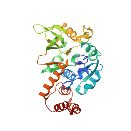
    The synthesis of deoxy-thymidine di-phosphate (dTDP)-L-rhamnose, an important component of the cell wall of many microorganisms, is a target for therapeutic intervention. The first enzyme in the dTDP-L-rhamnose biosynthetic pathway is glucose-1-phosphate thymidylyltransferase (RmlA). RmlA is inhibited by dTDP-L-rhamnose thereby regulating L-rhamnose production in bacteria. The structure of Pseudomonas aeruginosa RmlA has been solved to 1.66 A resolution. RmlA is a homotetramer, with the monomer consisting of three functional subdomains. The sugar binding and dimerization subdomains are unique to RmlA-like enzymes. The sequence of the core subdomain is found not only in sugar nucleotidyltransferases but also in other nucleotidyltransferases. The structures of five distinct enzyme substrate- product complexes reveal the enzyme mechanism that involves precise positioning of the nucleophile and activation of the electrophile. All the key residues are within the core subdomain, suggesting that the basic mechanism is found in many nucleotidyltransferases. The dTDP-L-rhamnose complex identifies how the protein is controlled by its natural inhibitor. This work provides a platform for the design of novel drugs against pathogenic bacteria.


  • Organizational Affiliation
    • The Centre for Biomolecular Sciences, The University, St Andrews, KY16 9ST, UK.

Macromolecule Content 

  • Total Structure Weight: 134.82 kDa 
  • Atom Count: 9,453 
  • Modeled Residue Count: 1,169 
  • Deposited Residue Count: 1,172 
  • Unique protein chains: 1

Macromolecules

Find similar proteins by:|  3D Structure
Entity ID: 1
MoleculeChains  Sequence LengthOrganismDetailsImage
GLUCOSE-1-PHOSPHATE THYMIDYLYLTRANSFERASE
A, B, C, D
293Pseudomonas aeruginosaMutation(s): 0 
EC: 2.7.7.24
UniProt
Find proteins for Q9HU22 (Pseudomonas aeruginosa (strain ATCC 15692 / DSM 22644 / CIP 104116 / JCM 14847 / LMG 12228 / 1C / PRS 101 / PAO1))
Explore Q9HU22 
Go to UniProtKB:  Q9HU22
Entity Groups
Sequence Clusters30% Identity50% Identity70% Identity90% Identity95% Identity100% Identity
UniProt GroupQ9HU22
Sequence Annotations
Expand
Reference Sequence

Small Molecules

Ligands 2 Unique
IDChains Name / Formula / InChI Key2D Diagram3D Interactions
TRH

Query on TRH


Download:Ideal Coordinates CCD File
F [auth A]
G [auth A]
J [auth B]
K [auth B]
M [auth C]
F [auth A],
G [auth A],
J [auth B],
K [auth B],
M [auth C],
N [auth C],
P [auth D],
Q [auth D]
2'-DEOXY-THYMIDINE-BETA-L-RHAMNOSE
C16 H26 N2 O15 P2
ZOSQFDVXNQFKBY-CGAXJHMRSA-N
SO4

Query on SO4


Download:Ideal Coordinates CCD File
E [auth A],
H [auth B],
I [auth B],
L [auth C],
O [auth D]
SULFATE ION
O4 S
QAOWNCQODCNURD-UHFFFAOYSA-L

Experimental Data & Validation

Experimental Data

  • Method: X-RAY DIFFRACTION
  • Resolution: 2.70 Å
  • R-Value Free:  0.230 (Depositor), 0.234 (DCC) 
  • R-Value Work:  0.204 (Depositor), 0.213 (DCC) 
  • R-Value Observed: 0.205 (Depositor) 
Space Group: P 21 21 21
Unit Cell:
Length ( Å )Angle ( ˚ )
a = 71.087α = 90
b = 138.557β = 90
c = 139.676γ = 90
Software Package:
Software NamePurpose
MOLREPphasing
REFMACrefinement
MOSFLMdata reduction
CCP4data scaling

Structure Validation

View Full Validation Report


Ligand Structure Quality Assessment 


Entry History 

Deposition Data

Revision History  (Full details and data files)

  • Version 1.0: 2000-12-27
    Type: Initial release
  • Version 1.1: 2008-04-27
    Changes: Version format compliance
  • Version 1.2: 2011-07-13
    Changes: Version format compliance
  • Version 1.3: 2018-03-07
    Changes: Data collection
  • Version 1.4: 2023-08-09
    Changes: Data collection, Database references, Derived calculations, Refinement description