1G0X | pdb_00001g0x

CRYSTAL STRUCTURE OF THE LIGAND BINDING DOMAIN OF LIR-1 (ILT2)


Experimental Data Snapshot

  • Method: X-RAY DIFFRACTION
  • Resolution: 2.10 Å
  • R-Value Free: 
    0.244 (Depositor) 
  • R-Value Work: 
    0.214 (Depositor) 
  • R-Value Observed: 
    0.214 (Depositor) 

wwPDB Validation 3D Report Full Report

Validation slider image for 1G0X

This is version 1.3 of the entry. See complete history

Literature

Crystal structure and ligand binding properties of the D1D2 region of the inhibitory receptor LIR-1 (ILT2).

Chapman, T.L.Heikema, A.P.West Jr., A.P.Bjorkman, P.J.

(2000) Immunity 13: 727-736

  • DOI: https://doi.org/10.1016/s1074-7613(00)00071-6
  • Primary Citation Related Structures: 
    1G0X

  • PubMed Abstract: 

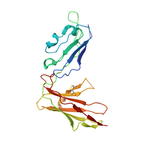
    LIR-1 is an inhibitory receptor that recognizes class I MHC molecules and the human cytomegalovirus class I homolog UL18. Here, we report the 2.1 A resolution crystal structure of the ligand binding portion of LIR-1 (domains 1 and 2 [D1D2]) and localize the binding region for UL18. LIR-1 D1D2 is composed of two immunoglobulin-like domains arranged at an acute angle to form a bent structure resembling the structures of natural killer inhibitory receptors (KIRs). The LIR-1 binding site comprises a portion of D1 distant from the interdomain hinge region that constitutes the KIR binding site, consistent with differences in LIR-1 and KIR recognition properties and functions.


  • Organizational Affiliation
    • Division of Biology 156-29, California Institute of Technology, Pasadena, CA 91125, USA.

Macromolecule Content 

  • Total Structure Weight: 22 kDa 
  • Atom Count: 1,757 
  • Modeled Residue Count: 192 
  • Deposited Residue Count: 197 
  • Unique protein chains: 1

Macromolecules

Find similar proteins by:|  3D Structure
Entity ID: 1
MoleculeChains  Sequence LengthOrganismDetailsImage
LEUCOCYTE IMMUNOGLOBULIN-LIKE RECEPTOR-1197Homo sapiensMutation(s): 0 
UniProt & NIH Common Fund Data Resources
Find proteins for Q8NHL6 (Homo sapiens)
Explore Q8NHL6 
Go to UniProtKB:  Q8NHL6
PHAROS:  Q8NHL6
Entity Groups
Sequence Clusters30% Identity50% Identity70% Identity90% Identity95% Identity100% Identity
UniProt GroupQ8NHL6
Sequence Annotations
Expand
Reference Sequence

Experimental Data & Validation

Experimental Data

  • Method: X-RAY DIFFRACTION
  • Resolution: 2.10 Å
  • R-Value Free:  0.244 (Depositor) 
  • R-Value Work:  0.214 (Depositor) 
  • R-Value Observed: 0.214 (Depositor) 
Space Group: P 41 21 2
Unit Cell:
Length ( Å )Angle ( ˚ )
a = 68.258α = 90
b = 68.258β = 90
c = 129.734γ = 90
Software Package:
Software NamePurpose
DENZOdata reduction
SCALEPACKdata scaling
AMoREphasing
CNSrefinement

Structure Validation

View Full Validation Report


Entry History 

Deposition Data

Revision History  (Full details and data files)

  • Version 1.0: 2001-01-17
    Type: Initial release
  • Version 1.1: 2008-04-27
    Changes: Version format compliance
  • Version 1.2: 2011-07-13
    Changes: Version format compliance
  • Version 1.3: 2024-11-06
    Changes: Data collection, Database references, Structure summary