1FZV | pdb_00001fzv

THE CRYSTAL STRUCTURE OF HUMAN PLACENTA GROWTH FACTOR-1 (PLGF-1), AN ANGIOGENIC PROTEIN AT 2.0A RESOLUTION


Experimental Data Snapshot

  • Method: X-RAY DIFFRACTION
  • Resolution: 2.00 Å
  • R-Value Free: 
    0.247 (Depositor) 
  • R-Value Work: 
    0.216 (Depositor) 
  • R-Value Observed: 
    0.216 (Depositor) 

wwPDB Validation 3D Report Full Report

Validation slider image for 1FZV

This is version 1.5 of the entry. See complete history

Literature

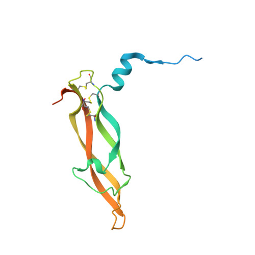
The crystal structure of human placenta growth factor-1 (PlGF-1), an angiogenic protein, at 2.0 A resolution.

Iyer, S.Leonidas, D.D.Swaminathan, G.J.Maglione, D.Battisti, M.Tucci, M.Persico, M.G.Acharya, K.R.

(2001) J Biological Chem 276: 12153-12161

  • DOI: https://doi.org/10.1074/jbc.M008055200
  • Primary Citation Related Structures: 
    1FZV

  • PubMed Abstract: 

    The angiogenic molecule placenta growth factor (PlGF) is a member of the cysteine-knot family of growth factors. In this study, a mature isoform of the human PlGF protein, PlGF-1, was crystallized as a homodimer in the crystallographic asymmetric unit, and its crystal structure was elucidated at 2.0 A resolution. The overall structure of PlGF-1 is similar to that of vascular endothelial growth factor (VEGF) with which it shares 42% amino acid sequence identity. Based on structural and biochemical data, we have mapped several important residues on the PlGF-1 molecule that are involved in recognition of the fms-like tyrosine kinase receptor (Flt-1, also known as VEGFR-1). We propose a model for the association of PlGF-1 and Flt-1 domain 2 with precise shape complementarity, consider the relevance of this assembly for PlGF-1 signal transduction, and provide a structural basis for altered specificity of this molecule.


  • Organizational Affiliation
    • Department of Biology and Biochemistry, University of Bath, Claverton Down, Bath BA2 7AY, United Kingdom.

Macromolecule Content 

  • Total Structure Weight: 29.98 kDa 
  • Atom Count: 1,694 
  • Modeled Residue Count: 200 
  • Deposited Residue Count: 264 
  • Unique protein chains: 1

Macromolecules

Find similar proteins by:|  3D Structure
Entity ID: 1
MoleculeChains  Sequence LengthOrganismDetailsImage
PLACENTA GROWTH FACTOR
A, B
132Homo sapiensMutation(s): 0 
UniProt & NIH Common Fund Data Resources
Find proteins for P49763 (Homo sapiens)
Explore P49763 
Go to UniProtKB:  P49763
PHAROS:  P49763
GTEx:  ENSG00000119630 
Entity Groups
Sequence Clusters30% Identity50% Identity70% Identity90% Identity95% Identity100% Identity
UniProt GroupP49763
Sequence Annotations
Expand
Reference Sequence

Small Molecules

Ligands 1 Unique
IDChains Name / Formula / InChI Key2D Diagram3D Interactions
MPD

Query on MPD


Download:Ideal Coordinates CCD File
C [auth A],
D [auth B]
(4S)-2-METHYL-2,4-PENTANEDIOL
C6 H14 O2
SVTBMSDMJJWYQN-YFKPBYRVSA-N

Experimental Data & Validation

Experimental Data

  • Method: X-RAY DIFFRACTION
  • Resolution: 2.00 Å
  • R-Value Free:  0.247 (Depositor) 
  • R-Value Work:  0.216 (Depositor) 
  • R-Value Observed: 0.216 (Depositor) 
Space Group: P 43
Unit Cell:
Length ( Å )Angle ( ˚ )
a = 62.6α = 90
b = 62.6β = 90
c = 84.107γ = 90
Software Package:
Software NamePurpose
DENZOdata reduction
SCALEPACKdata scaling
AMoREphasing
CNSrefinement

Structure Validation

View Full Validation Report


Entry History 

Deposition Data

Revision History  (Full details and data files)

  • Version 1.0: 2001-05-09
    Type: Initial release
  • Version 1.1: 2008-04-27
    Changes: Version format compliance
  • Version 1.2: 2011-07-13
    Changes: Version format compliance
  • Version 1.3: 2018-01-31
    Changes: Experimental preparation
  • Version 1.4: 2022-12-21
    Changes: Database references, Derived calculations
  • Version 1.5: 2024-10-16
    Changes: Data collection, Structure summary