Calf spleen purine nucleoside phosphorylase: structure of its ternary complex with an N(7)-acycloguanosine inhibitor and a phosphate anion.
Luic, M., Koellner, G., Shugar, D., Saenger, W., Bzowska, A.(2001) Acta Crystallogr D Biol Crystallogr 57: 30-36
- PubMed: 11134924 Search on PubMed
- DOI: https://doi.org/10.1107/s0907444900014402
- Primary Citation Related Structures:
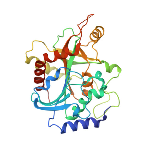
1FXU - PubMed Abstract:
The calf spleen purine nucleoside phosphorylase (PNP) ternary complex with an N(7)-acycloguanosine inhibitor and a phosphate ion has been crystallized in the cubic space group P2(1)3, with unit-cell parameter a = 94.11 A and one monomer per asymmetric unit. X-ray diffraction data were collected using synchrotron radiation (Station X31, EMBL Outstation, DESY, Hamburg). The crystal structure was refined to a resolution of 2.2 A and R and R(free) values of 17.5 and 24.5%, respectively. The acyclonucleoside inhibitor is bound in the active site in an inverted ('upside-down') orientation of the purine base compared with natural substrates. The side chain of Asp243 forms two hydrogen bonds with the base ring: N(delta) donates a hydrogen to N(3) and O(delta) accepts a hydrogen from the guanine N(2)-amino group. N(1)--H of the base is hydrogen bonded to O(epsilon) of Glu201, while N(9) accepts a hydrogen bond from Thr242 O(gamma). In addition, a water molecule (W417) bridges the N(2)-amino group of the base and O(epsilon) of Glu201. In the phosphate-binding site, a phosphate ion is bound to Ser33, His64, Arg84, His86, Ala116 and Ser220. The acyclic chain of the N(7)-acycloguanosine inhibitor is in a folded conformation and together with a water molecule (W388) occupies the pentose-binding site, with possible hydrogen bonds to Tyr88 O(eta) and His257 N(delta 1). This new binding mode fully accounts for the previously observed substrate properties of 7-beta-D-ribofuranosides of hypoxanthine and guanine. It also provides a new starting point for the design of inhibitors of PNP for therapeutic and other applications.
- Rudjer Boskovic Institute, Bijenicka 54, 10000 Zagreb, Croatia.
Organizational Affiliation: