1FVI | pdb_00001fvi

CRYSTAL STRUCTURE OF CHLORELLA VIRUS DNA LIGASE-ADENYLATE


Experimental Data Snapshot

  • Method: X-RAY DIFFRACTION
  • Resolution: 2.00 Å
  • R-Value Free: 
    0.270 (Depositor) 
  • R-Value Work: 
    0.190 (Depositor) 

wwPDB Validation 3D Report Full Report

Validation slider image for 1FVI

This is version 1.4 of the entry. See complete history

Literature

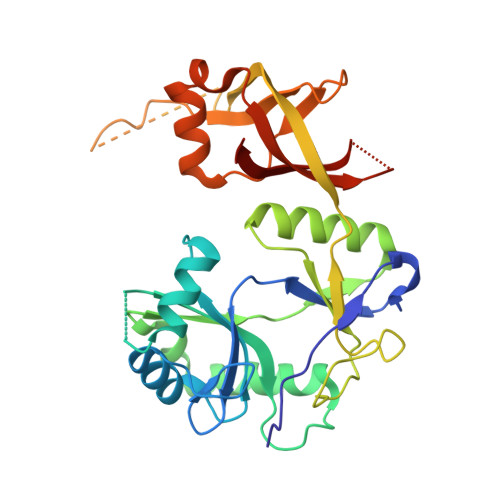
Crystal structure of eukaryotic DNA ligase-adenylate illuminates the mechanism of nick sensing and strand joining.

Odell, M.Sriskanda, V.Shuman, S.Nikolov, D.B.

(2000) Mol Cell 6: 1183-1193

  • DOI: https://doi.org/10.1016/s1097-2765(00)00115-5
  • Primary Citation Related Structures: 
    1FVI

  • PubMed Abstract: 

    Chlorella virus DNA ligase is the smallest eukaryotic ATP-dependent ligase known; it has an intrinsic nick-sensing function and suffices for yeast cell growth. Here, we report the 2.0 A crystal structure of the covalent ligase-AMP reaction intermediate. The conformation of the adenosine nucleoside and contacts between the enzyme and the ribose sugar have undergone a significant change compared to complexes of T7 ligase with ATP or mRNA capping enzyme with GTP. The conformational switch allows the 3' OH of AMP to coordinate directly the 5' PO(4) of the nick. The structure explains why nick sensing is restricted to adenylated ligase and why the 5' phosphate is required for DNA binding. We identify a metal binding site on ligase-adenylate and propose a mechanism of nick recognition and catalysis supported by mutational data.


  • Organizational Affiliation
    • Molecular Biology Program and Cellular Biochemistry and Biophysics Program Sloan-Kettering Institute, New York, NY 10021, USA.

Macromolecule Content 

  • Total Structure Weight: 34.57 kDa 
  • Atom Count: 2,430 
  • Modeled Residue Count: 264 
  • Deposited Residue Count: 297 
  • Unique protein chains: 1

Macromolecules

Find similar proteins by:|  3D Structure
Entity ID: 1
MoleculeChains  Sequence LengthOrganismDetailsImage
CHLORELLA VIRUS DNA LIGASE-ADENYLATE297Chlorella virusMutation(s): 1 
EC: 6.5.1
UniProt
Find proteins for A0ACD6B8E9 (Paramecium bursaria Chlorella virus AR158)
Explore A0ACD6B8E9 
Go to UniProtKB:  A0ACD6B8E9
Entity Groups
Sequence Clusters30% Identity50% Identity70% Identity90% Identity95% Identity100% Identity
UniProt GroupA0ACD6B8E9
Sequence Annotations
Expand
Reference Sequence

Small Molecules

Ligands 2 Unique
IDChains Name / Formula / InChI Key2D Diagram3D Interactions
AMP

Query on AMP


Download:Ideal Coordinates CCD File
C [auth A]ADENOSINE MONOPHOSPHATE
C10 H14 N5 O7 P
UDMBCSSLTHHNCD-KQYNXXCUSA-N
SO4

Query on SO4


Download:Ideal Coordinates CCD File
B [auth A]SULFATE ION
O4 S
QAOWNCQODCNURD-UHFFFAOYSA-L

Experimental Data & Validation

Experimental Data

  • Method: X-RAY DIFFRACTION
  • Resolution: 2.00 Å
  • R-Value Free:  0.270 (Depositor) 
  • R-Value Work:  0.190 (Depositor) 
Space Group: P 21 21 21
Unit Cell:
Length ( Å )Angle ( ˚ )
a = 48.17α = 90
b = 58.44β = 90
c = 101.79γ = 90
Software Package:
Software NamePurpose
MLPHAREphasing
X-PLORrefinement
DENZOdata reduction
SCALEPACKdata scaling

Structure Validation

View Full Validation Report


Entry History 

Deposition Data

Revision History  (Full details and data files)

  • Version 1.0: 2000-11-22
    Type: Initial release
  • Version 1.1: 2008-04-27
    Changes: Version format compliance
  • Version 1.2: 2011-07-13
    Changes: Version format compliance
  • Version 1.3: 2021-11-03
    Changes: Database references, Derived calculations
  • Version 1.4: 2024-11-20
    Changes: Data collection, Structure summary