1FRX | pdb_00001frx

STRUCTURE AND PROPERTIES OF C20S FDI MUTANT


Experimental Data Snapshot

  • Method: X-RAY DIFFRACTION
  • Resolution: 2.50 Å
  • R-Value Work: 
    0.229 (Depositor) 
  • R-Value Observed: 
    0.229 (Depositor) 

wwPDB Validation 3D Report Full Report

Validation slider image for 1FRX

This is version 1.3 of the entry. See complete history

Literature

Site-directed mutagenesis of Azotobacter vinelandii ferredoxin I: cysteine ligation of the [4Fe-4S] cluster with protein rearrangement is preferred over serine ligation.

Shen, B.Jollie, D.R.Diller, T.C.Stout, C.D.Stephens, P.J.Burgess, B.K.

(1995) Proc Natl Acad Sci U S A 92: 10064-10068

  • DOI: https://doi.org/10.1073/pnas.92.22.10064
  • Primary Citation Related Structures: 
    1FRX

  • PubMed Abstract: 

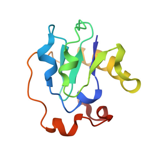
    The [4Fe-4S] cluster of Azotobacter vinelandii ferredoxin I receives three of its four ligands from a Cys-Xaa-Xaa-Cys-Xaa-Xaa-Cys sequence at positions 39-45 while the fourth ligand, Cys20, is provided by a distal portion of the sequence. Previously we reported that the site-directed mutation of Cys20 to Ala (C20A protein) resulted in the formation of a new [4Fe-4S] cluster that obtained its fourth ligand from Cys24, a free cysteine in the native structure. That ligand exchange required significant protein rearrangement. Here we report the conversion of Cys20 to Ser (C20S protein), which gives the protein the opportunity either to retain the native structure and use the Ser20 O gamma as a ligand or to rearrange and use Cys24. X-ray crystallography demonstrates that the cluster does not use the Ser20 O gamma as a ligand; rather it rearranges to use Cys24. In the C20S protein the [4Fe-4S] cluster has altered stability and redox properties relative to either C20A or the native protein.


  • Organizational Affiliation
    • Department of Molecular Biology and Biochemistry, University of California, Irvine 92717, USA.

Macromolecule Content 

  • Total Structure Weight: 12.69 kDa 
  • Atom Count: 878 
  • Modeled Residue Count: 106 
  • Deposited Residue Count: 106 
  • Unique protein chains: 1

Macromolecules

Find similar proteins by:|  3D Structure
Entity ID: 1
MoleculeChains  Sequence LengthOrganismDetailsImage
FERREDOXIN106Azotobacter vinelandiiMutation(s): 0 
UniProt
Find proteins for P00214 (Azotobacter vinelandii)
Explore P00214 
Go to UniProtKB:  P00214
Entity Groups
Sequence Clusters30% Identity50% Identity70% Identity90% Identity95% Identity100% Identity
UniProt GroupP00214
Sequence Annotations
Expand
Reference Sequence

Small Molecules

Ligands 2 Unique
IDChains Name / Formula / InChI Key2D Diagram3D Interactions
SF4

Query on SF4


Download:Ideal Coordinates CCD File
B [auth A]IRON/SULFUR CLUSTER
Fe4 S4
LJBDFODJNLIPKO-UHFFFAOYSA-N
F3S

Query on F3S


Download:Ideal Coordinates CCD File
C [auth A]FE3-S4 CLUSTER
Fe3 S4
FCXHZBQOKRZXKS-UHFFFAOYSA-N

Experimental Data & Validation

Experimental Data

  • Method: X-RAY DIFFRACTION
  • Resolution: 2.50 Å
  • R-Value Work:  0.229 (Depositor) 
  • R-Value Observed: 0.229 (Depositor) 
Space Group: P 41 21 2
Unit Cell:
Length ( Å )Angle ( ˚ )
a = 55.25α = 90
b = 55.25β = 90
c = 95.45γ = 90
Software Package:
Software NamePurpose
X-PLORmodel building
X-PLORrefinement
X-PLORphasing

Structure Validation

View Full Validation Report


Entry History 

Deposition Data

  • Released Date: 1994-08-31 
  • Deposition Author(s): Stout, C.D.

Revision History  (Full details and data files)

  • Version 1.0: 1994-08-31
    Type: Initial release
  • Version 1.1: 2008-03-24
    Changes: Version format compliance
  • Version 1.2: 2011-07-13
    Changes: Version format compliance
  • Version 1.3: 2024-02-07
    Changes: Data collection, Database references, Derived calculations, Other