1FPS | pdb_00001fps

CRYSTAL STRUCTURE OF RECOMBINANT FARNESYL DIPHOSPHATE SYNTHASE AT 2.6 ANGSTROMS RESOLUTION


Experimental Data Snapshot

  • Method: X-RAY DIFFRACTION
  • Resolution: 2.60 Å
  • R-Value Work: 
    0.194 (Depositor) 
  • R-Value Observed: 
    0.194 (Depositor) 

wwPDB Validation 3D Report Full Report

Validation slider image for 1FPS

This is version 1.3 of the entry. See complete history

Literature

Crystal structure of recombinant farnesyl diphosphate synthase at 2.6-A resolution.

Tarshis, L.C.Yan, M.Poulter, C.D.Sacchettini, J.C.

(1994) Biochemistry 33: 10871-10877

  • DOI: https://doi.org/10.1021/bi00202a004
  • Primary Citation Related Structures: 
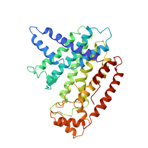
    1FPS

  • PubMed Abstract: 

    The synthesis of farnesyl diphosphate (FPP), a key intermediate in the isoprenoid biosynthetic pathway required for the synthesis of cholesterol and in the formation of prenylated proteins, is catalyzed by the enzyme farnesyl diphosphate synthase (FPS). The crystal structure of avian recombinant FPS, the first three-dimensional structure for any prenyltransferase, was determined to 2.6-A resolution. The enzyme exhibits a novel fold composed entirely of alpha-helices joined by connecting loops. The enzyme's most prominent structural feature is the arrangement of 10 core helices around a large central cavity. Two aspartate-rich sequences that are highly conserved among the isoprenyl diphosphate synthase family of prenyltransferases, and are essential for enzymatic activity, were found on opposite walls of this cavity, with the aspartate side chains approximately 12 A apart and facing each other. The location and metal ion binding properties of these sequences suggest that the conserved aspartate residues participate in substrate binding of catalysis.


  • Organizational Affiliation
    • Department of Biochemistry, Albert Einstein College of Medicine, Bronx, New York 10461.

Macromolecule Content 

  • Total Structure Weight: 40.02 kDa 
  • Atom Count: 2,894 
  • Modeled Residue Count: 348 
  • Deposited Residue Count: 348 
  • Unique protein chains: 1

Macromolecules

Find similar proteins by:|  3D Structure
Entity ID: 1
MoleculeChains  Sequence LengthOrganismDetailsImage
FARNESYL DIPHOSPHATE SYNTHASE348Gallus gallusMutation(s): 0 
EC: 2.5.1.10 (PDB Primary Data), 2.5.1.1 (UniProt)
UniProt
Find proteins for P08836 (Gallus gallus)
Explore P08836 
Go to UniProtKB:  P08836
Entity Groups
Sequence Clusters30% Identity50% Identity70% Identity90% Identity95% Identity100% Identity
UniProt GroupP08836
Sequence Annotations
Expand
Reference Sequence

Experimental Data & Validation

Experimental Data

  • Method: X-RAY DIFFRACTION
  • Resolution: 2.60 Å
  • R-Value Work:  0.194 (Depositor) 
  • R-Value Observed: 0.194 (Depositor) 
Space Group: I 41 2 2
Unit Cell:
Length ( Å )Angle ( ˚ )
a = 88.6α = 90
b = 88.6β = 90
c = 276.1γ = 90
Software Package:
Software NamePurpose
X-PLORmodel building
X-PLORrefinement
X-PLORphasing

Structure Validation

View Full Validation Report


Entry History 

Deposition Data

Revision History  (Full details and data files)

  • Version 1.0: 1995-07-10
    Type: Initial release
  • Version 1.1: 2008-03-24
    Changes: Version format compliance
  • Version 1.2: 2011-07-13
    Changes: Derived calculations, Version format compliance
  • Version 1.3: 2024-02-07
    Changes: Data collection, Database references, Other