1FOS | pdb_00001fos

TWO HUMAN C-FOS:C-JUN:DNA COMPLEXES


Experimental Data Snapshot

  • Method: X-RAY DIFFRACTION
  • Resolution: 3.05 Å
  • R-Value Free: 
    0.321 (Depositor) 
  • R-Value Work: 
    0.230 (Depositor) 
  • R-Value Observed: 
    0.230 (Depositor) 

wwPDB Validation 3D Report Full Report

Validation slider image for 1FOS

This is version 1.4 of the entry. See complete history

Literature

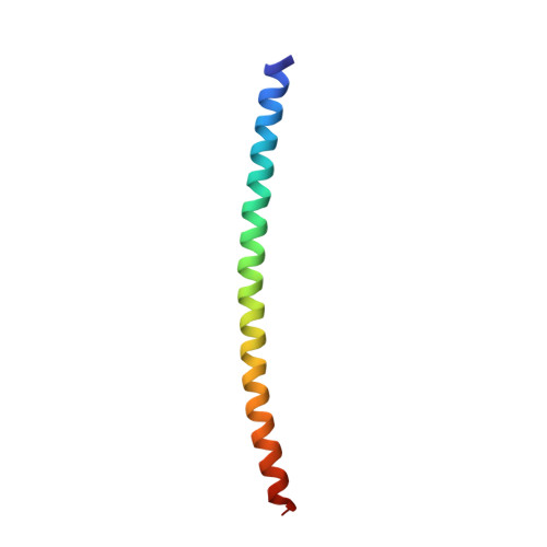
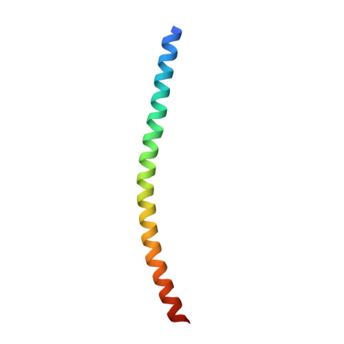
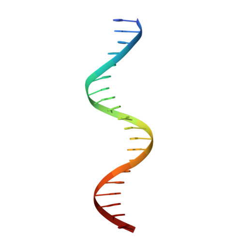
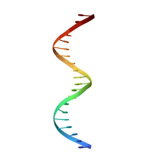
Crystal structure of the heterodimeric bZIP transcription factor c-Fos-c-Jun bound to DNA.

Glover, J.N.Harrison, S.C.

(1995) Nature 373: 257-261

  • DOI: https://doi.org/10.1038/373257a0
  • Primary Citation Related Structures: 
    1FOS

  • PubMed Abstract: 

    The Fos and Jun families of eukaryotic transcription factors heterodimerize to form complexes capable of binding 5'-TGAGTCA-3' DNA elements. We have determined the X-ray crystal structure of a heterodimer of the bZIP regions of c-Fos and c-Jun bound to DNA. Both subunits form continuous alpha-helices. The carboxy-terminal regions form an asymmetric coiled-coil, and the amino-terminal regions make base-specific contacts with DNA in the major groove. Comparison of the two crystallographically distinct protein-DNA complexes show that the coiled-coil is flexibly joined to the basic regions and that the Fos-Jun heterodimer does not recognize the asymmetric 5'-TGAGTCA-3' recognition element in a unique orientation. There is an extensive network of electrostatic interactions between subunits within the coiled-coil, consistent with proposals that these interactions determine preferential formation of the heterodimer over either of the homodimers.


  • Organizational Affiliation
    • Howard Hughes Medical Institute, Harvard University, Cambridge, Massachusetts 02138.

Macromolecule Content 

  • Total Structure Weight: 53.93 kDa 
  • Atom Count: 3,480 
  • Modeled Residue Count: 314 
  • Deposited Residue Count: 328 
  • Unique protein chains: 2
  • Unique nucleic acid chains: 2

Macromolecules


Find similar proteins by:|  3D Structure
Entity ID: 3
MoleculeChains  Sequence LengthOrganismDetailsImage
P55-C-FOS PROTO-ONCOGENE PROTEIN
E, G
62Homo sapiensMutation(s): 1 
UniProt & NIH Common Fund Data Resources
Find proteins for P01100 (Homo sapiens)
Explore P01100 
Go to UniProtKB:  P01100
PHAROS:  P01100
GTEx:  ENSG00000170345 
Entity Groups
Sequence Clusters30% Identity50% Identity70% Identity90% Identity95% Identity100% Identity
UniProt GroupP01100
Sequence Annotations
Expand
Reference Sequence
Find similar proteins by:|  3D Structure
Entity ID: 4
MoleculeChains  Sequence LengthOrganismDetailsImage
C-JUN PROTO-ONCOGENE PROTEIN
F, H
62Homo sapiensMutation(s): 1 
UniProt & NIH Common Fund Data Resources
Find proteins for P05412 (Homo sapiens)
Explore P05412 
Go to UniProtKB:  P05412
PHAROS:  P05412
GTEx:  ENSG00000177606 
Entity Groups
Sequence Clusters30% Identity50% Identity70% Identity90% Identity95% Identity100% Identity
UniProt GroupP05412
Sequence Annotations
Expand
Reference Sequence
Find similar nucleic acids by:  Sequence
Entity ID: 1
MoleculeChains LengthOrganismImage
DNA (5'-D(*AP*AP*TP*GP*GP*AP*TP*GP*AP*GP*TP*CP*AP*TP*AP*GP*GP*AP*GP*A)-3')
A, C
20N/A
Sequence Annotations
Expand
Reference Sequence
Find similar nucleic acids by:  Sequence
Entity ID: 2
MoleculeChains LengthOrganismImage
DNA (5'-D(*TP*TP*CP*TP*CP*CP*TP*AP*TP*GP*AP*CP*TP*CP*AP*TP*CP*CP*AP*T)-3')
B, D
20N/A
Sequence Annotations
Expand
Reference Sequence

Experimental Data & Validation

Experimental Data

  • Method: X-RAY DIFFRACTION
  • Resolution: 3.05 Å
  • R-Value Free:  0.321 (Depositor) 
  • R-Value Work:  0.230 (Depositor) 
  • R-Value Observed: 0.230 (Depositor) 
Space Group: P 21 21 2
Unit Cell:
Length ( Å )Angle ( ˚ )
a = 241.1α = 90
b = 48.69β = 90
c = 66γ = 90
Software Package:
Software NamePurpose
X-PLORrefinement
XDSdata reduction

Structure Validation

View Full Validation Report


Entry History 

Deposition Data

Revision History  (Full details and data files)

  • Version 1.0: 1995-07-10
    Type: Initial release
  • Version 1.1: 2008-05-22
    Changes: Version format compliance
  • Version 1.2: 2011-07-13
    Changes: Version format compliance
  • Version 1.3: 2021-11-03
    Changes: Database references
  • Version 1.4: 2024-02-07
    Changes: Data collection