1FLJ | pdb_00001flj

CRYSTAL STRUCTURE OF S-GLUTATHIOLATED CARBONIC ANHYDRASE III


Experimental Data Snapshot

  • Method: X-RAY DIFFRACTION
  • Resolution: 1.80 Å
  • R-Value Free: 
    0.213 (Depositor) 
  • R-Value Work: 
    0.155 (Depositor) 
  • R-Value Observed: 
    0.155 (Depositor) 

wwPDB Validation 3D Report Full Report

Validation slider image for 1FLJ

This is version 1.5 of the entry. See complete history

Literature

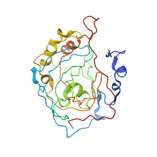
Crystal structure of S-glutathiolated carbonic anhydrase III.

Mallis, R.J.Poland, B.W.Chatterjee, T.K.Fisher, R.A.Darmawan, S.Honzatko, R.B.Thomas, J.A.

(2000) FEBS Lett 482: 237-241

  • DOI: https://doi.org/10.1016/s0014-5793(00)02022-6
  • Primary Citation Related Structures: 
    1FLJ

  • PubMed Abstract: 

    S-Glutathiolation of carbonic anhydrase III (CAIII) occurs rapidly in hepatocytes under oxidative stress. The crystal structure of the S-glutathiolated CAIII from rat liver reveals covalent adducts on cysteines 183 and 188. Electrostatic charge and steric contacts at each modification site inversely correlate with the relative rates of reactivity of these cysteines toward glutathione (GSH). Diffuse electron density associated with the GSH adducts suggests a lack of preferred bonding interactions between CAIII and the glutathionyl moieties. Hence, the GSH adducts are available for binding by a protein capable of reducing this mixed disulfide. These properties are consistent with the participation of CAIII in the protection/recovery from the damaging effects of oxidative agents.


  • Organizational Affiliation
    • Department of Biochemistry, Biophysics and Molecular Biology, Iowa State University, Ames, IA 50011, USA.

Macromolecule Content 

  • Total Structure Weight: 30.05 kDa 
  • Atom Count: 2,365 
  • Modeled Residue Count: 260 
  • Deposited Residue Count: 260 
  • Unique protein chains: 1

Macromolecules

Find similar proteins by:|  3D Structure
Entity ID: 1
MoleculeChains  Sequence LengthOrganismDetailsImage
CARBONIC ANHYDRASE III260Rattus norvegicusMutation(s): 0 
EC: 4.2.1.1
UniProt
Find proteins for P14141 (Rattus norvegicus)
Explore P14141 
Go to UniProtKB:  P14141
Entity Groups
Sequence Clusters30% Identity50% Identity70% Identity90% Identity95% Identity100% Identity
UniProt GroupP14141
Sequence Annotations
Expand
Reference Sequence

Biologically Interesting Molecules (External Reference) 

1 Unique
Entity ID: 3
IDChains NameType/Class2D Diagram3D Interactions
PRD_002593 (GSH)
Query on PRD_002593
C [auth A],
D [auth A]
GlutathionePeptide-like / Oxidation-reduction

Experimental Data & Validation

Experimental Data

  • Method: X-RAY DIFFRACTION
  • Resolution: 1.80 Å
  • R-Value Free:  0.213 (Depositor) 
  • R-Value Work:  0.155 (Depositor) 
  • R-Value Observed: 0.155 (Depositor) 
Space Group: P 1 21 1
Unit Cell:
Length ( Å )Angle ( ˚ )
a = 41.25α = 90
b = 44.73β = 99.73
c = 66.91γ = 90
Software Package:
Software NamePurpose
X-PLORmodel building
X-PLORrefinement
X-GENdata reduction
X-GENdata scaling
X-PLORphasing

Structure Validation

View Full Validation Report


Entry History 

Deposition Data

Revision History  (Full details and data files)

  • Version 1.0: 2000-09-04
    Type: Initial release
  • Version 1.1: 2008-04-27
    Changes: Version format compliance
  • Version 1.2: 2011-07-13
    Changes: Version format compliance
  • Version 1.3: 2011-12-21
    Changes: Non-polymer description
  • Version 1.4: 2017-10-04
    Changes: Refinement description
  • Version 1.5: 2025-03-26
    Changes: Data collection, Database references, Derived calculations, Structure summary